Guide To Identifying A Port Wine Stain
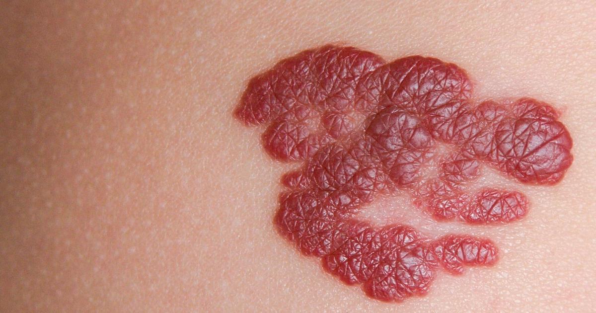
Medically known as a naevus flammeus, a port-wine stain is a skin discoloration that occurs due to a malformation of capillaries. Approximately three out of every one thousand children are born with port-wine stains, and the capillary malformation that produces them is linked to a genetic mutation that takes place before a baby is born. The mutation is not hereditary, so it is not passed down to future generations. For reasons not yet understood, females are twice as likely to develop port-wine stains as males. The birthmarks can be easily diagnosed with a visual inspection of the skin, and laser therapy is usually the preferred treatment method. Patients may also choose to use concealing makeup.
The major characteristics of port-wine stains are discussed below.
Color And Texture

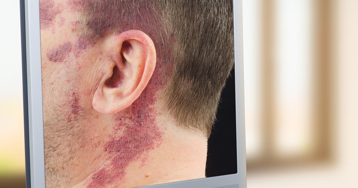
At birth, port-wine stains are typically light pink or reddish. As a child grows, the color of the birthmark often darkens with time and usually reaches a deep purple by the time the child enters adulthood. The texture of this particular birthmark tends to be smooth from birth and throughout childhood. Adults who have port-wine stains might occasionally notice the texture of their birthmark becomes thicker with age, and many patients in their forties and above have observed it takes on a cobbled texture with ridges and tiny bumps elevated above the skin's surface. While these are often normal changes, any changes in a birthmark should be examined by a dermatologist to make sure they are benign. In addition, patients should always protect their port-wine stain from potential darkening due to sun damage. Applying sunscreen to the birthmark daily is recommended.
Read about how size and location play into identifying a port-wine stain next.
Size And Location

Port-wine stains can vary in size. The smallest stains may be just a few millimeters in diameter, and some patients may have larger stains that cover almost half their face. As a child grows, the size of their port-wine stain will likely increase in proportion to their growth. While port-wine stains most frequently affect only one side of the body, cases of port-wine stains on both sides of the body have been observed. Port-wine stains may develop in any location, but they are most commonly seen on the face, scalp, neck, arms, legs, and upper chest. An estimated sixty-five percent of all port-wine stains are found on the head and neck. Port-wine stains located on the face or neck respond especially well to treatment with a pulsed dye laser.
Uncover details on the types of port-wine stains to look out for next.
Types Of Port-Wine Stains

'Port-wine stain' is a non-medical term often used by the public to refer to any birthmark doctors would classify as a capillary vascular malformation. One of the most common types of port-wine stains (capillary vascular malformations) in this group is known as naevus simplex. This particular birthmark affects up to forty percent of newborns, and it is a light pink patch that typically appears at the nape of the neck or on the eyelids. It can also appear in between the eyebrows, and this presentation is referred to as an 'angel's kiss.' Naevus simplex lesions become increasingly red when a child cries, holds their breath, strains, or engages in vigorous activity, and the color of the lesions can also be influenced by the ambient temperature. Naevus simplex patches often become lighter with age, and the patches are not painful or itchy. Unlike true port-wine stains (nevus flammeus), naevus simplex lesions at locations other than the nape of the neck often fade within the first two years of a child's life. Those located at the nape of the neck fade at a slower rate, and fifty percent of lesions in this area could persist indefinitely. Since the nape of the neck is often covered by hair, the lesions might not be of cosmetic concern to the patient.
Discover more information about port-wine stains now.
Do They Bleed

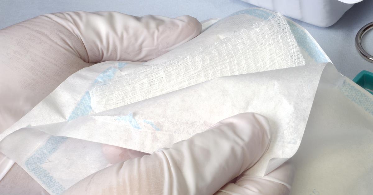
Capillary vascular malformations, including port-wine stains, are generally more prone to bleeding after minor trauma than other areas of skin. Scratching or picking the birthmark could cause bleeding, and patients need to be especially mindful of protecting the site from injury. If bleeding does occur, firm pressure should be applied to the site of the bleeding for at least ten minutes. The area should be cleaned with soap and water, and some patients might wish to apply antibiotic cream. A sterile bandage should be placed over the injury, and patients will need to change it at least once a day, though it should be changed more frequently if it gets wet or dirty. As the area heals, it is important to watch for possible signs of infection. For example, individuals should get medical attention if they notice any discharge of pus, swelling, or warmth at the site of the birthmark.
Learn about other potential symptoms linked to port-wine stains now.
Do They Cause Other Symptoms?

While most port-wine stains are harmless and do not cause any additional symptoms, patients who have these birthmarks might occasionally develop blood vessel blisters such as papules and pyogenic granulomas. The blisters can bleed easily, and doctors recommend treating them with laser therapy, freezing methods (cryotherapy), or surgery. The tissue underneath a port-wine stain might enlarge, and this could result in soft tissue hypertrophy, a condition that occurs most often with port-wine stains near the lips. In addition, port-wine stains are sometimes associated with more serious medical conditions. Patients who have a port-wine stain around their eye should have their eye pressure monitored. Individuals with a port-wine stain on their eye, forehead, or scalp should be evaluated for Sturge-Weber syndrome, a rare condition that may cause seizures and learning difficulties. The presence of a large port-wine stain on the arms or legs is sometimes associated with Klippel-Trenaunay syndrome. This syndrome causes the affected limb to grow longer than the unaffected side. If a leg is affected and is less than two centimeters longer than the unaffected leg, patients may be given specialized insoles to wear in their shoes. If more than two centimeters of growth occurs, surgical interventions could be recommended.
