Complications Linked To Muscular Dystrophy
Muscular dystrophy is the overarching term for a group of diseases that lead the body to become progressively weaker and lose muscle mass. The condition is caused by abnormal gene mutations that interfere with the body's ability to produce proteins that help form healthy muscles. Because there are multiple types of muscular dystrophy, the complications and age of onset vary. The symptoms also vary depending on what muscle groups are affected.
The most common variety begins showing symptoms in childhood, but other types don't cause symptoms until an individual reaches adulthood. While muscular dystrophy doesn't have a cure, there are medications and physical therapy options that can help slow the disease and manage overall symptoms.
Heart Problems
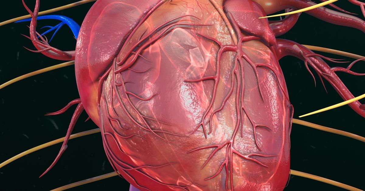
Research has shown multiple types of muscular dystrophy include cardiac dysfunction. Depending on the case and the underlying subtype, some people might experience heart problems as the main symptom of the illness. Because muscular dystrophy tends to be a rare issue, and each subtype is also rare, cardiologists might struggle to provide a diagnosis for the heart issues. Until recently, researchers weren't aware of how often cardiomyopathy causes death in muscular dystrophy patients. However, recent advances in respiratory care have improved survival rates. It's important to detect muscular dystrophy-related heart problems early.
While it might not be possible to stop the heart issues from developing, instituting heart-healthy therapies can sometimes help to slow the progression of the disease. Echocardiography and electrocardiography tests are recommended for screening patients for heart disease. There has also been some research indicating cardiovascular magnetic resonance screening can reveal early heart involvement in muscular dystrophy patients. The most common types of this condition including the heart are those that affect dystrophin proteins, like Becker and Duchenne muscular dystrophy. Other types that show involvement are Emery-Dreifuss, limb-girdle, and myotonic.
Trouble Walking
Muscular dystrophy can cause trouble walking, though the amount of leg involvement varies depending on the type. Some patients might progress to the full use of a wheelchair, while others may only need mobility devices like braces. The condition can also cause difficulty standing up. Duchenne muscular dystrophy is the most commonly recognized subtype, and it tends to show symptoms in childhood. Patients with this form usually have such progressive muscle weakness they need to use a wheelchair before they reach their teens.
Because of how rapidly the disease progresses and how it impairs the heart and lungs, children with this disease don't usually live past their twenties. Becker muscular dystrophy has similar symptoms to Duchenne, but it also occurs between eleven and twenty-five years old, and the severity is less pronounced. Patients with Becker muscular dystrophy might walk on their toes, fall frequently, experience muscle cramps and spasms, and struggle to get up from the floor.
Breathing Difficulties
Breathing difficulties are common in individuals with muscular dystrophy, though the level of involvement varies from case to case. Problems with breathing typically occur because there's a progressive weakening of the respiratory muscles. The diaphragm is an important muscle for respiration. It is located just underneath the lungs and assists in inhaling. When the diaphragm weakens in patients with muscular dystrophy, they aren't able to inhale as deeply, which leads to a reduction in oxygen and a decrease in lung function. There are other muscles in the abdomen that help with exhaling. Abdominal weakness can make it difficult to exhale fully, which means the lungs aren't cleared of carbon dioxide.
Another issue is weakening of the muscles that make up the upper respiratory tract. The muscles in the throat and nose are necessary for air intake. They can collapse when sleeping, which makes muscular dystrophy patients more likely to have breathing issues while they sleep. These patients might be more hurt by illnesses like pneumonia because the weakened respiratory muscles make it difficult to cough up phlegm. If an individual with muscular dystrophy has scoliosis, it can affect how the chest wall is structured and lead to breathing issues.
Scoliosis
Scoliosis is a condition characterized by the abnormal sideways curving of the spine. While the condition can occur by itself, it's also often a complication of muscular dystrophy. Many of these patients have scoliosis, and in this case, the condition is caused by progressive weakness in the muscles that support the spinal column. Most patients don't develop scoliosis until after the disease has progressed enough so they are no longer able to walk or stand. If this is combined with progressive tilting of the patient's pelvis, it can be difficult to sit because there's an uneven distribution of weight between the buttocks.
As with other complications, the presence of scoliosis varies widely depending on the type of muscular dystrophy and symptom progression. Some patients with congenital muscular dystrophy might have scoliosis from birth. With Ullrich congenital muscular dystrophy, scoliosis tends to develop within the first ten years of the patient's life. Seventy to ninety percent of teenagers with Duchenne muscular dystrophy develop scoliosis. The condition has also been seen in the Becker, limb-girdle, and facioscapulohumeral subtypes.
Contractures
Contractures, seen in many types of muscular dystrophy, occur when the joints become fixed and lose part or all of their range of motion. This can lead to pain and discomfort in the surrounding muscles. Contractures are found in the fingers, wrists, elbows, feet, hips, and knees. Many patients with Duchenne muscular dystrophy experience contractures in their legs as the disease causes progressive muscle wasting. There are several ways to help minimize or slow the progression of contractures in patients with muscular dystrophy.
One way is to use range-of-motion exercises, which are physical therapy exercises that help retain and increase the joint's range of motion. Typically, patients should perform these according to a regular schedule. Doing this can keep the tendons from undergoing premature shortening, which delays the onset of contractures.