Signs Of A Branchial Cleft Cyst And How It's Diagnosed
A branchial cleft cyst is an open space or pouch in the tissue around the neck or shoulder, most commonly found between the neck and collarbone. Although it may not be noticed until late childhood or early adulthood, the branchial cleft cyst is a common congenital defect. It is caused when structures around the neck develop abnormally within the womb, leaving a space instead of solid tissue. The condition is often diagnosed when the cyst fills with mucus, causing it to bulge and form a lump. A patient may mistake the cyst for a cancerous mass due to the unusual nature of this growth.
Fluid Drainage From The Neck
A branchial cleft cyst can exist for years without being noticed. As a child's body matures, the sinuses can drain more effectively during a cold or another illness. When a child or young adult has a branchial cleft cyst, mucus caused by the infection can drain into the cyst, causing it to swell. In most cases, the cyst is interior and feels like a lump under the skin. In other cases, there may be a small hole, known as a fistula, leading from the cyst to the outer skin. The cyst becomes noticeable when there is fluid drainage from the neck. Although this symptom may be troubling, it is only a problem if the cyst itself becomes infected. In some cases, surgery can be done to remove the cyst and close the fistula.
Presence Of A Skin Tag Or Dimple
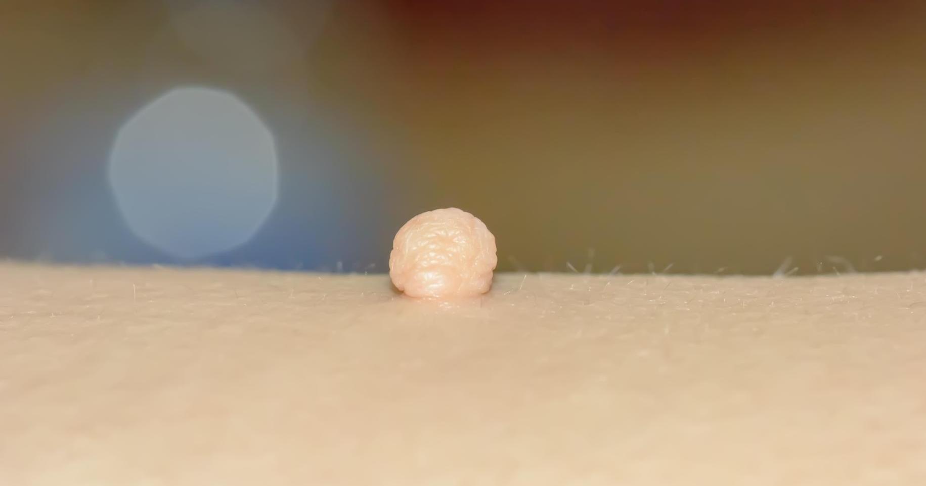
The branchial cleft cyst is the result of incomplete growth of tissue in the womb. This may also result in some kind of marking on the outer skin at the site of the cyst, such as a skin tag or dimple. As opposed to a fistula, which allows drainage from the cyst to the outer skin, a skin tag or dimple indicates the growth process in the womb was more complete. Because these skin markings can be found in other places on the body, a skin tag or dimple does not necessarily indicate a cyst. It is when the cyst swells that skin tags and dimples can help confirm a diagnosis.
Swelling Or Tenderness Around The Neck
Swelling or tenderness around the neck is often the first sign that brings a patient with a branchial cleft cyst to the doctor. These cysts are normally found on one side of the neck, though they do sometimes present as pairs. This swelling is often associated with a cold or another infection that requires the sinuses to drain. The cyst fills with mucus, becoming swollen and uncomfortable. Depending on the size and placement, swelling can interfere with swallowing. When the cyst is infected, its walls become thicker making it even more noticeable. If a doctor suspects infection, they will prescribe antibiotics. In more extreme cases, the doctor will order incision and drainage of the cyst.
Diagnostic Imaging Tests
Diagnostic imaging tests are an important tool in diagnosing a branchial cleft cyst. The primary concern is making certain the lump is a relatively harmless cyst and not something more serious like a cancerous growth. The most common tests ordered are ultrasound, CT, and MRI scans. Of these three, the MRI gives the clearest image of the cyst. When doctors examine the scans, they hope to see a liquid-filled structure and not a solid lump. A cyst will usually have a well-defined, smooth oval shape without the irregularities of a cancerous growth. Another indicator of a cyst as opposed to cancer is the lack of independent blood supply to the structure.
Fine Needle Aspiration
Diagnostic scans are a preferable means of examining a suspected branchial cleft cyst because they are non-invasive. However, a doctor may order fine needle aspiration for a more definite diagnosis. By drawing some of the fluid out of the cyst, the doctor can determine the nature of the object and the extent of any infection. Clear liquid indicates a normal branchial cleft cyst. If the liquid is yellow and pus-like, it indicates an infection in the cyst. The test will also examine what kind of cells are present in the object. A cyst is lined with squamous skin cells, the type of cells lining the hollow organs of the body. Cancerous cells indicate a growing tumor.