How To Spot And Diagnose X-Linked Retinoschisis
Decline In Vision

One of the first symptoms of X-linked retinoschisis is a lack of visual acuity that can't be corrected with glasses. In cases where the condition is progressive, patients may experience a decline in vision that leads to legal blindness. When the retinal layers become separated, cystic macular lesions form between them. These are similar to blisters. The lesions might increase the thickness of the retina, which will make an individual's vision loss worse. However, they can be treated with different medications. Since the condition affects the retina's ability to develop, symptoms tend to present early in childhood. Children should have vision tests at six months old, three years old, prior to first grade, and every two years throughout their school years.
Learn about how X-linked retinoschisis is diagnosed now.
Electroretinography

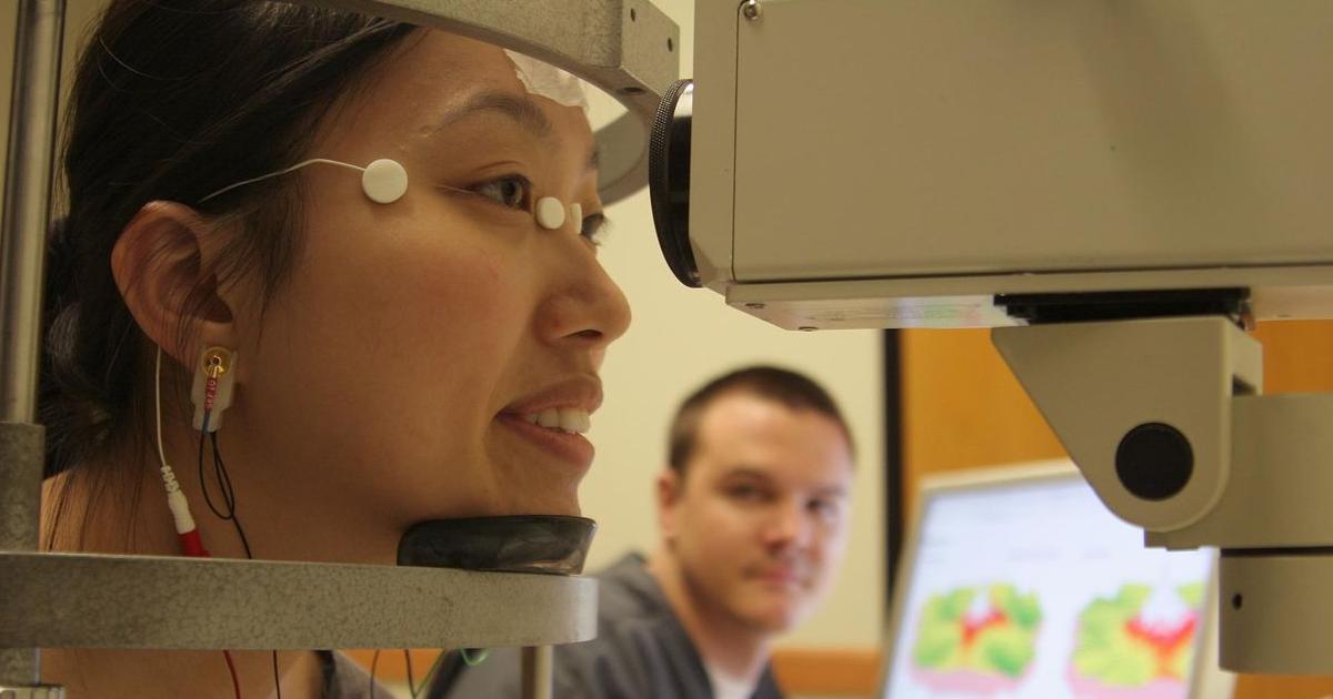
Electroretinography is a diagnostic eye test that determines how well the retina functions. The retina has multiple layers of specialized cells. The rods and cones are photoreceptor cells that detect light. There are also ganglion cells to transmit visual data to the brain. Electroretinography detects electrical signals given off by the photoreceptors, plus electrical signals from the bipolar and Muller cells located between the ganglion and photoreceptor cells. When the readings are abnormal, they can help an ophthalmologist determine what retinal abnormalities are causing vision loss. The test is done by placing an electrode on a patient's cornea to measure the eye's electrical responses to light.
Keep reading for more information on how X-linked retinoschisis is diagnosed now.
