What Is Astrocytoma?
An astrocytoma is a type of brain tumor that develops in the specialized brain and spinal cord cells referred to as neurons or astrocytes. Tumors that initially form from the glia cells in the brain are referred to as gliomas, and astrocytes are a type of glial cell in the brain. The most prevalent form of glial tumors are astrocytomas, which comprise half of all diagnosed primary brain tumors. Astrocytomas occur more often in men than in women, and they tend to manifest after the fourth decade of life. The causes of astrocytomas are not currently known, but immunologic abnormalities, genetic abnormalities, stress, diet, exposure to ionizing radiation, certain chemicals, and ultraviolet rays are factors with a contributory role in its pathophysiology. Astrocytomas occur more often in individuals affected by Turcot syndrome, Ollier's disease, Li-Fraumeni syndrome, and neurofibromatosis type-I tuberous sclerosis.
Grading And Types

Astrocytomas are typed and graded based on the different features seen on pathology slides according to how normal or abnormal the cells appear. The different grades of astrocytomas have varying behavior patterns. Pineal astrocytic tumors are a type of astrocytoma that can be any grade and develop in the pineal gland in the cerebellum. Brain stem gliomas are a rare type of astrocytoma that can be any grade and develop in the area where the spinal cord enters the brain. Subependymal giant cell astrocytomas and pilocytic astrocytomas are considered grade I and are most prominent among children. Diffuse astrocytomas are considered to be grade II and slow-growing, but they have the potential to grow into nearby tissue. Anaplastic astrocytomas are considered to be grade III tumors that are fast-growing and spread quickly to neighboring tissue. Glioblastomas are considered grade IV tumors that grow rapidly into nearby tissues and are difficult to treat because they are a blend of various cancer cell types. Over half of all cases of astrocytomas are diagnosed as glioblastomas.
Locations Found

Some types of astrocytomas are characterized by the location in the brain where they are found. Astrocytomas that begin in the tissues that make up the cerebellum are referred to as cerebellar astrocytomas. The cerebellum, a region inside the brain located near the base of the skull, is responsible for the coordination of balance and muscle movements. An optic pathway glioma is a type of astrocytoma that begins in the cells around or in the optic nerve, which is responsible for the transmission of visual information from the eyes to the vision center in the brain. A hypothalamic glioma and thalamic glioma are a type of astrocytoma that begins in the region of the brain referred to as the thalamus. The thalamus is a relay center for an individual's movements and is responsible for the identification of sensations like pain, touch, and temperature changes. The hypothalamus sits just underneath the thalamus and functions to regulate an individual's body temperature, appetite, sleep, and hormone function. A tectal glioma is a type of astrocytoma that forms in the brain stem roof, which is responsible for an individual's heart rate, blood pressure, and breathing rate.
Common Symptoms

Several common symptoms occur in individuals with an astrocytoma. The severity and range of symptoms are dependent upon the location of the tumor in the brain. A slow-growing grade I or grade II astrocytoma may not manifest with noticeable symptoms since the brain can adapt to the slow-growing tumor being present over time. When symptoms do manifest, they are caused by growing pressure inside the skull on different parts of the brain or from the location of the tumor itself. Twitching and numbness in an individual's arm, leg, or face can be a manifestation of an astrocytoma, particularly when seizures are involved. Headaches that are persistent and severe, double or blurred vision, nausea and vomiting, appetite loss, mood and personality changes, changes in the ability to learn and think, gradual onset speech difficulties, and general weakness are all symptoms that are indicative of an astrocytoma. Should the astrocytoma develop in the cells located in the spinal cord, symptoms would include clumsiness in the legs, arms, and gait, along with bladder and bowel control problems.
Diagnostic Measures

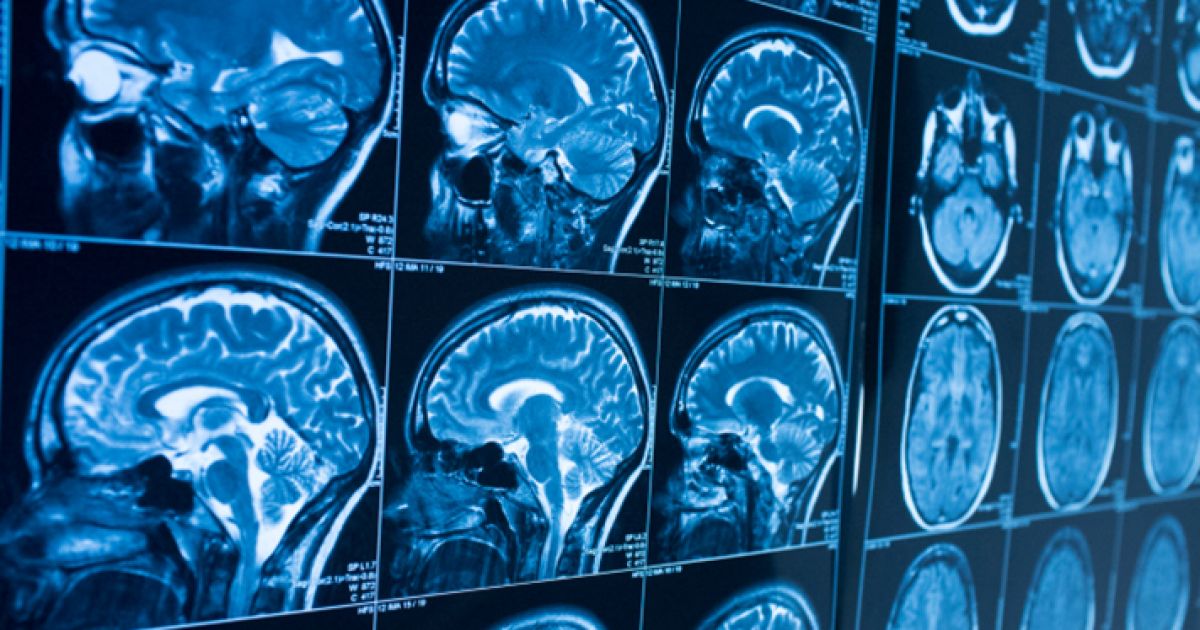
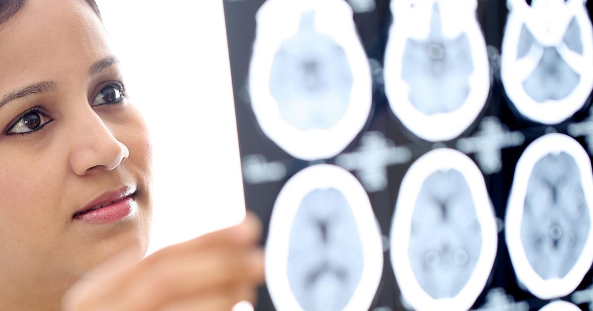
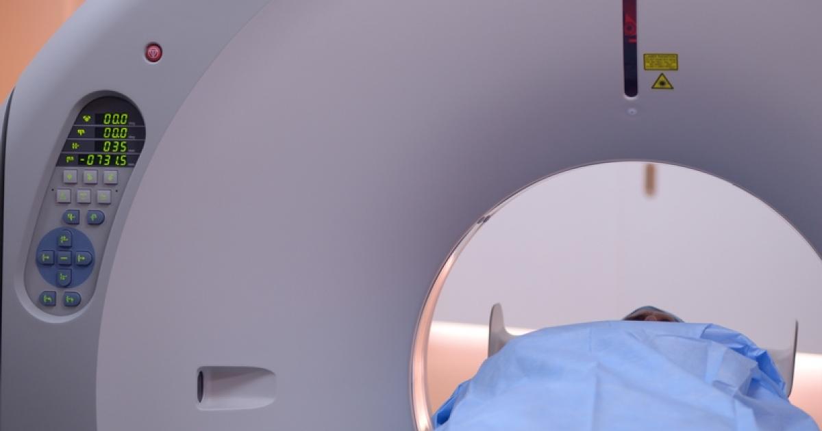
Certain diagnostic measures must be taken to identify the cause of numerous neurologically-related symptoms that may indicate the presence of an astrocytoma or other brain tumors. Careful patient history, thorough clinical evaluation, characteristic physical findings, and several specialized tests are utilized in the diagnosis of an astrocytoma. Specialized tests used to identify an astrocytoma as the underlying problem include diagnostic imaging techniques such as CT and MRI scans. The specificity and sensitivity of these imaging techniques can be enhanced when intravenous contrast medium is administered to the patient during the scans. Careful administration of pre-medication can help physicians overcome a patient's allergy to dye in cases where contrast scans are necessary. Neuroimaging tests help a physician evaluate the size, location, and other characteristics of a patient's tumor. Once a tumor is confirmed, a brain tissue biopsy procedure is carried out to obtain a sample that is sent to the laboratory for a microscopic examination of the malignant cells to identify the tumor grade and type.
Treatment And Prognosis

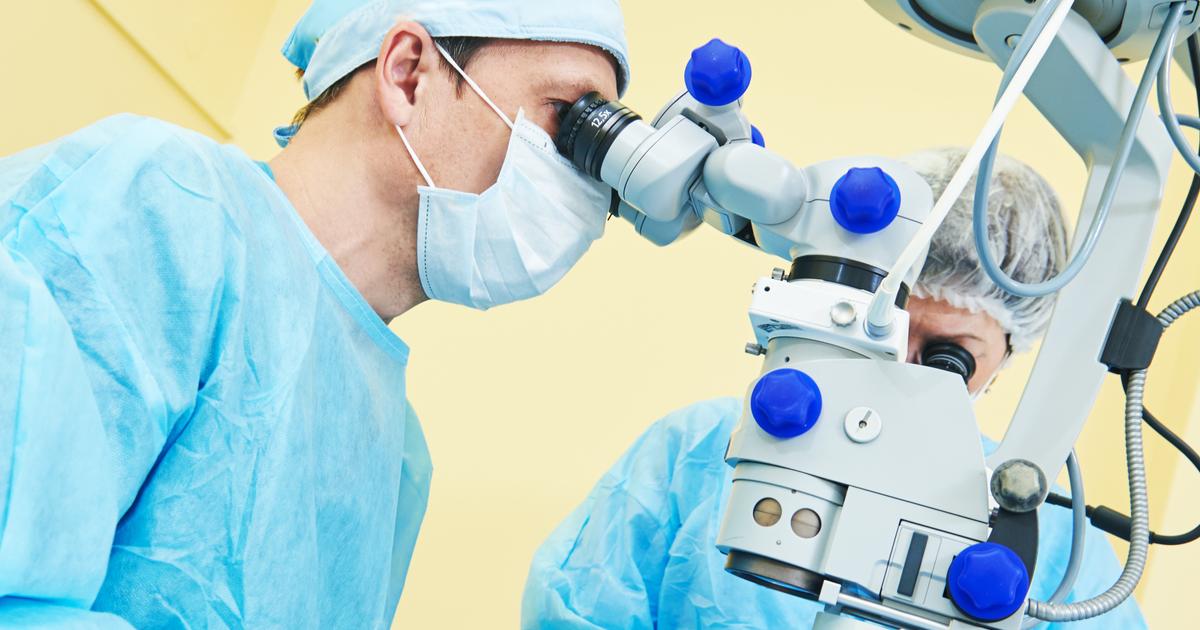
The grade and type of an astrocytoma are what determines the appropriate treatment and patient prognosis. Grade I astrocytomas can usually be cured with excision surgery if they are not in an inaccessible part of the brain. Grade II astrocytomas are also treated using excision surgery but may require the removal of additional tissues with different important functions or may need additional treatment using chemotherapy or radiation. Malignant grade III and grade IV astrocytomas are treated with aggressive excision surgery and chemotherapy that is concurrent with up to six weeks of fractionated radiation therapy. Neurologists can use brain mapping techniques and technology before and during the surgical procedure to minimize the risk of complications and injury of tissues that surround the tumor. In many cases of grade III and IV astrocytomas, the entire tumor is not able to be removed with a surgical excision procedure. However, the surgery does decrease the bulk of the tumor and relieve the pressure being put on adjacent brain tissues. After chemotherapy and radiation therapy of grade III and IV astrocytomas, diligent monitoring with surveillance MRI scans is essential for early detection of a reoccurrence.
