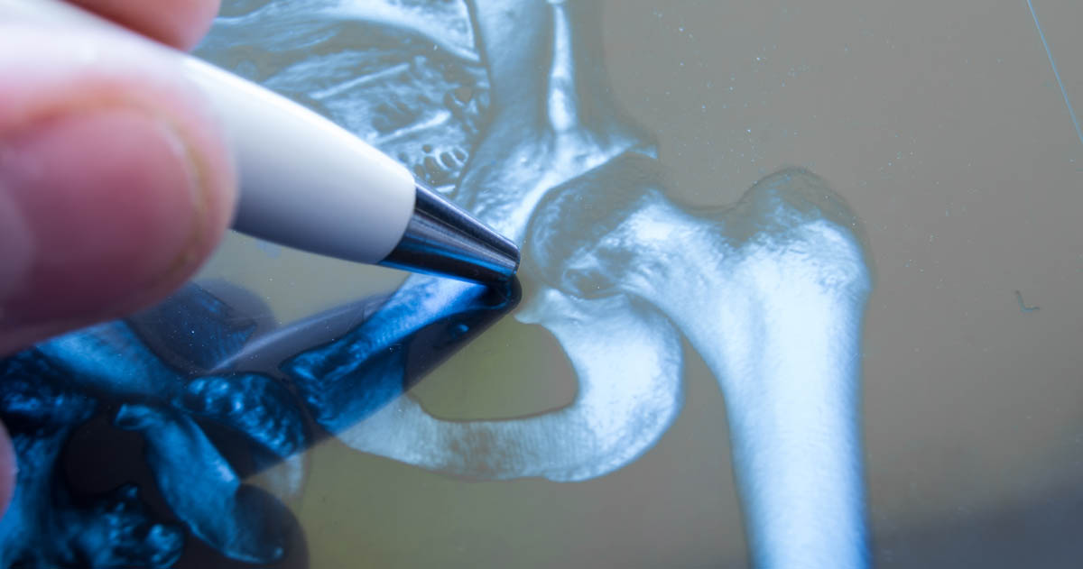
How Does An X-Ray Work?
X-rays are some of the most common imaging tools in medicine. They can be used to detect and diagnose a great deal of different bodily issues, from broken bones to blocked blood vessels and cancer. But how do x-rays work? You might have heard they use radiation to create their images. Being nervous about the use of radiation in medicine is natural. But x-ray technology has become safer and safer over the years, with qualified technicians and researchers helping to minimize the risks. X-rays are an essential part of being a medical professional, and they're also an essential element to diagnose many conditions. It's crucial to have a full understanding of what an x-ray is, and how the technology works.
What Is An X-Ray?

An x-ray is a type of electromagnetic radiation, similar to the light we can see visibly. However, unlike light, x-rays have higher amounts of energy, which allows them to pass through the majority of objects, including your body. A medical x-ray is used to generate pictures of structures and tissues throughout the body. When an x-ray that travels through the body passes through a detector on the patient's other side, an image forms to represent the 'shadows' created by objects found inside the body. There are many different x-ray detectors. One type is photographic film, and others can be used for digital image production. When people capture x-ray images through this process, they are referred to as radiographs.
Continue reading to learn about how x-rays work.
How Does It Work?

How does an x-ray work? First, the patient will be positioned so whatever part of their body the doctor needs to view will be located between a source of x-rays and an x-ray detector. When the technician turns on the machine, x-rays travel through the person's body. The rays become absorbed in different ways, due to the different densities of the tissues in the body. Bones can absorb x-rays much more easily than other tissues in the body, which causes high contrast images to appear on the x-ray machine, and structures made of bone will appear to be more white than other tissues in the body. Meanwhile, x-rays have an easier time traveling through tissues like muscle and fat, along with the lungs and other cavities filled with air. Because the x-rays are not absorbed entirely, these tissues and structures appear in different gray shades upon a radiograph.
Continue reading to discover the different uses for x-rays.
Different Uses For X-Rays

There are lots of different uses in the medical field, but the two main ones are diagnostic and therapeutic. Diagnostic methods seek to diagnose a problem, while therapeutic methods seek to treat a problem. X-ray radiography is the most common form of x-ray and is used to detect abnormal masses, tumors, bone fractures, pneumonia, and foreign objects in the body. Mammography is a radiograph done of a breast to gauge whether a woman has breast cancer or not.
A CT scan is a combination of computer processing and traditional x-ray technology. This combination generates cross-sectional body images, which are then turned into a three-dimensional x-ray, as opposed to a conventional two-dimensional x-ray. Fluoroscopy is the use of a fluorescent screen and x-rays to receive images of real-time movement occurring inside the body. X-rays are also used in cancer treatment as a type of radiation therapy, where their high-energy radiation can destroy cancerous cells through damage to their DNA. Radiation doses for cancer treatments tend to be much higher than radiation doses for simple imaging.
Continue reading to learn about the safety of x-rays.
Is It Safe To Use?

As with most medical technology, there are some risks. However, there are so many diagnostic benefits that the risks are minimal in comparison. An x-ray scan has the potential to diagnose life-threatening conditions such as infections, bone cancer, and blocked blood vessels. But x-rays do create ionizing radiation, which can harm a person's living tissue, and the risk of harm increases with each subsequent exposure. With that said, risks of cancer development due to x-ray exposure are minimal. Pregnant women can be x-rayed without significant risk to their child as long as their pelvis and abdomen are not being imaged. If the pelvis and abdomen must be imaged, doctors prefer to use methods without radiation. Children have a higher risk of cancer development than adults because of their sensitivity to ionizing radiation. It's vital for the doctor or technician to adjust their machine settings to accommodate children.
Continue reading to reveal other uses for the machines themselves.
Other Uses For X-Ray Machines

What are some other uses for x-ray machines? Nearly every airport in the world has some kind of x-ray security system installed, which scans all baggage to make sure no dangerous items have been packed. In recent years, full-body scans have been implemented for additional security. Another lesser-known use for x-ray machines is when an art historian detects whether an image has been placed over an existing piece. X-ray technology helps to reveal counterfeit art, and it can even assist art historians in restoring the images found below the surface coat of paint.
In the medical field, researchers are currently working on new ways to reduce the radiation dosage, improve image resolutions, and enhance the contrast methods and materials. Different medical fields are the focus of various studies, with many advancements focused on CT technology and mammography.
