Causes And Risk Factors For A Cavernous Malformation
A cavernous malformation is a condition where an individual develops abnormally shaped blood vessels that cause issues in the spinal cord and brain. The blood vessel malformations can range in diameter from two millimeters to several centimeters and have a shape that resembles a small mulberry. Symptoms of a cavernous malformation include severe headache, vomiting, speech difficulties, vision loss, balance difficulties, nausea, numbness on one side of the body, and double vision.
Most cases of cavernous malformations are a single, idiopathic occurrence without the involvement of any genetic factors. However, around one-fifth of all individuals affected by cavernous malformations have an inherited or familial type of the disorder. When symptoms of a cavernous malformation emerge in an individual, MRI scans and genetic tests are performed to diagnose the disorder. Treatment for cerebral malformations is individualized and may include observation, medications, and surgery.
Genetic Mutations
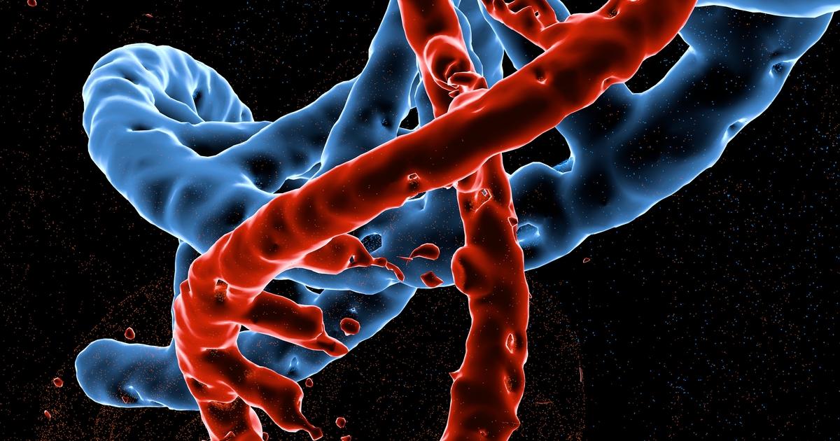
An individual affected by certain genetic mutations in their DNA may develop cavernous malformations as a complication of their genetic predisposition. Genetic mutations that cause an individual to develop cavernous malformations are inherited from parents in an autosomal dominant fashion. Around twenty percent of all cases of cavernous malformations are the result of an inherited genetic mutation. Mutations in the CCM3 gene are what produce the most severe form of cavernous malformation, and affected individuals tend to experience a hemorrhage at an early age.
Other mutations that cause the development of cavernous malformations include those in the CCM1 and CCM2 genes. These genes play critical roles in the maintenance and development of an individual's blood vessels. A mutation in one of these genes causes malformed blood vessels to develop in the patient's brain. These malformed blood vessels do not have good structural integrity in the junction between the cells that make up the blood vessels, allowing blood to leak into the brain easily.
Focal Brain Radiation Therapy
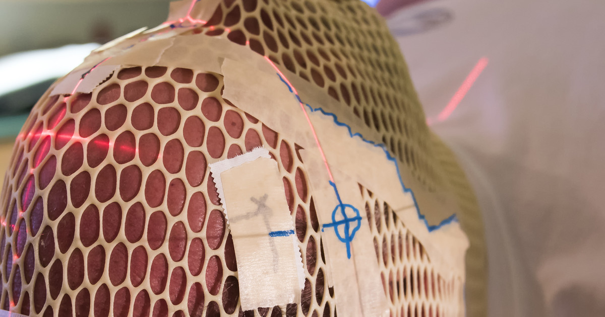
An individual who has undergone focal brain radiation therapy is at a greater risk of developing cavernous malformations than someone who has not. Focal brain radiation therapy is a method of treatment for certain types of cancerous and benign tumors that develop in the brain. Radiation therapy uses potent beams of energy that contain x-rays or particles to destroy or damage the DNA of cancerous cells. However, these beams can also affect the DNA of healthy cells surrounding the tissue being treated.
When healthy cells are affected by radiation beams, mutations can occur as a result of the treatment or the body's attempt at repairing the damage from the treatment. A cavernous malformation can develop due to a somatic mutation that occurs in a single cell damaged by focal brain radiation therapy. Somatic mutation is a change in the DNA of a single cell that can be passed on to daughter cells when the cell undergoes cell division. These mutated cells can continue to reproduce and cause the development of a cavernous malformation. The average duration between radiation therapy and the diagnosis of cavernous malformation is around twelve years.
Trauma

An individual who experiences head trauma is more likely to be diagnosed with a cavernous malformation than those who do not. Head trauma does not cause an individual to develop a cavernous malformation but is what produces the discovery and subsequent diagnosis of a cavernous malformation in many cases. Trauma to the head can cause the fragile blood vessels that make up the cavernous malformation to burst and produce life-threatening bleeding in the brain. Head trauma has the potential to cause a hemorrhagic stroke in an individual who has a cavernous malformation.
When trauma to the head is evaluated with the use of diagnostic imaging tests such as an MRI or CT scan, any existing or ruptured cavernous malformations will be detected. Cavernous malformations can actually produce brain trauma when they grow large enough to take up space inside of the skull other tissues should be occupying. Micro-bleeding from the cavernous malformation in the brain can cause pressure to build up and compress tissues of the brain that leads to a traumatic brain injury. Facial trauma and neck trauma have also been implicated in a brain injury precipitated from a ruptured cavernous malformation upon impact.
Spinal Injury

An individual who has experienced a spinal injury is at a higher risk of having a cavernous malformation than others. Instead of a spinal injury causing the development of a cavernous malformation, the cavernous malformations can cause an individual to experience a spinal injury. This risk factor is a less prevalent one, and cavernous malformations are typically found on the imaging performed to evaluate the patient's spinal injury. Only between three and five percent of all cavernous malformations are found in a location within the spinal cord.
Half of all cavernous malformations that occur in the spinal cord are found in the thoracic section, forty percent are found in the cervical section, and ten percent are found in the conus. A spinal injury can occur in an individual who has a cavernous malformation in their spinal cord because of its growth occupancy of space, significant hemorrhage, and repeated micro-bleeding. Unlike cavernous malformations that develop in the brain, cavernous malformations that occur in the spinal column can cause an individual to experience a spinal injury that can result in progressive myelopathy and partial or complete paraplegia.
Ethnicity And Race

An individual who is of a particular ethnicity and race has an increased risk of developing cavernous malformations than individuals of other ethnicities and races. The prevalence of cavernous malformations among the general population is between 0.4 percent to 0.8 percent, even though they are the most prevalent malformation of a vascular nature. The prevalence of cavernous malformations is equal among males and females in the population.
In the United States, over half of all Hispanic individuals diagnosed with cavernous malformations are affected by a familial form of the disease. However, only ten to twenty percent of Caucasian individuals affected by a cavernous malformation is diagnosed with a familial form of the disease. The reason behind this prevalence of familial cavernous malformation disease in individuals of Hispanic ethnicity over other races is not known, and few studies have been carried out to investigate this particular characteristic or part of the disease etiology.
