Causes And Risk Factors For A Cavernous Malformation
A cavernous malformation is a condition where an individual develops abnormally shaped blood vessels that cause issues in the spinal cord and brain. The blood vessel malformations can range in diameter from two millimeters to several centimeters and have a shape that resembles a small mulberry. Symptoms of a cavernous malformation include severe headache, vomiting, speech difficulties, vision loss, balance difficulties, nausea, numbness on one side of the body, and double vision.
Most cases of cavernous malformations are a single, idiopathic occurrence without the involvement of any genetic factors. However, around one-fifth of all individuals affected by cavernous malformations have an inherited or familial type of the disorder. When symptoms of a cavernous malformation emerge in an individual, MRI scans and genetic tests are performed to diagnose the disorder. Treatment for cerebral malformations is individualized and may include observation, medications, and surgery.
Genetic Mutations

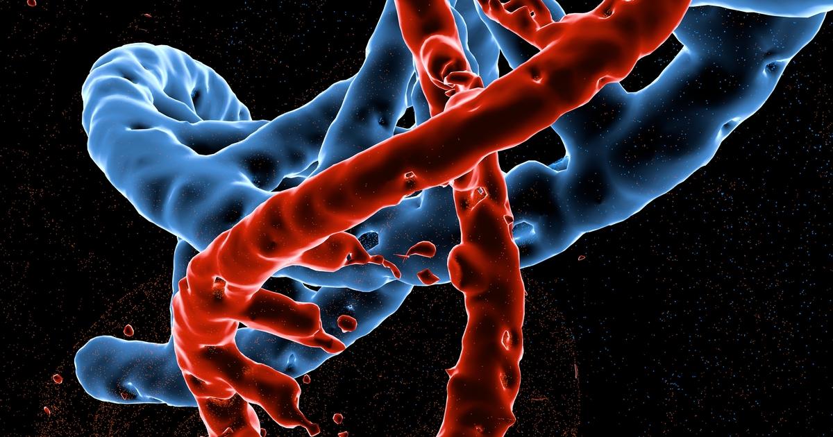
An individual affected by certain genetic mutations in their DNA may develop cavernous malformations as a complication of their genetic predisposition. Genetic mutations that cause an individual to develop cavernous malformations are inherited from parents in an autosomal dominant fashion. Around twenty percent of all cases of cavernous malformations are the result of an inherited genetic mutation. Mutations in the CCM3 gene are what produce the most severe form of cavernous malformation, and affected individuals tend to experience a hemorrhage at an early age.
Other mutations that cause the development of cavernous malformations include those in the CCM1 and CCM2 genes. These genes play critical roles in the maintenance and development of an individual's blood vessels. A mutation in one of these genes causes malformed blood vessels to develop in the patient's brain. These malformed blood vessels do not have good structural integrity in the junction between the cells that make up the blood vessels, allowing blood to leak into the brain easily.
Focal Brain Radiation Therapy

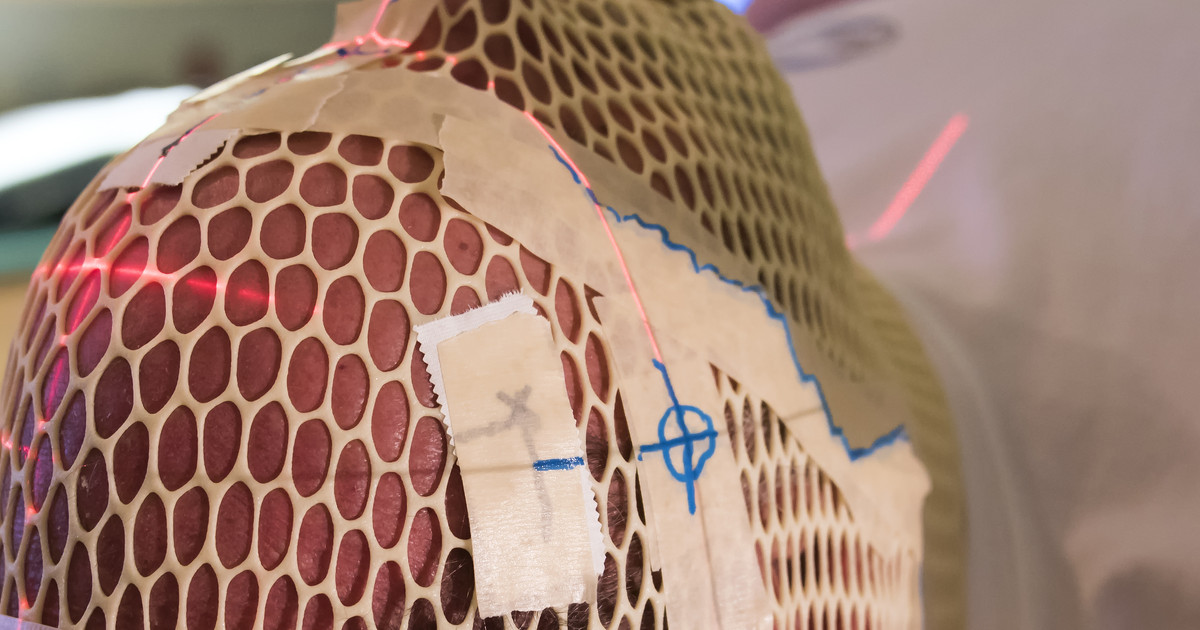
An individual who has undergone focal brain radiation therapy is at a greater risk of developing cavernous malformations than someone who has not. Focal brain radiation therapy is a method of treatment for certain types of cancerous and benign tumors that develop in the brain. Radiation therapy uses potent beams of energy that contain x-rays or particles to destroy or damage the DNA of cancerous cells. However, these beams can also affect the DNA of healthy cells surrounding the tissue being treated.
When healthy cells are affected by radiation beams, mutations can occur as a result of the treatment or the body's attempt at repairing the damage from the treatment. A cavernous malformation can develop due to a somatic mutation that occurs in a single cell damaged by focal brain radiation therapy. Somatic mutation is a change in the DNA of a single cell that can be passed on to daughter cells when the cell undergoes cell division. These mutated cells can continue to reproduce and cause the development of a cavernous malformation. The average duration between radiation therapy and the diagnosis of cavernous malformation is around twelve years.
