How To Diagnose And Treat Schwannomas
When a tumor develops in the peripheral nervous system or the cranial nerves within the Schwann cells, it is called a schwannoma. Schwann cells are responsible for the production of the protective myelin sheath around the axons of nerves. Most often, schwannomas are benign or noncancerous, and in the cases where a schwannoma is cancerous, it will be called a soft tissue sarcoma. One of the most common areas for a schwannoma to form is on the nerve connecting the inner ear to the brain. This is called an acoustic neuroma or a vestibular schwannoma. One of the most common areas where cancerous schwannomas occur is on the sciatic nerve of the leg. Schwannomas do not develop anywhere in the brain or the spinal cord. Symptoms will manifest when the schwannoma has grown large enough to put pressure on the nerves near it. The location of the tumor will be the main influence on what kind of symptoms an individual will experience.
Learn about how schwannomas are diagnosed and treated now.
Electromyogram
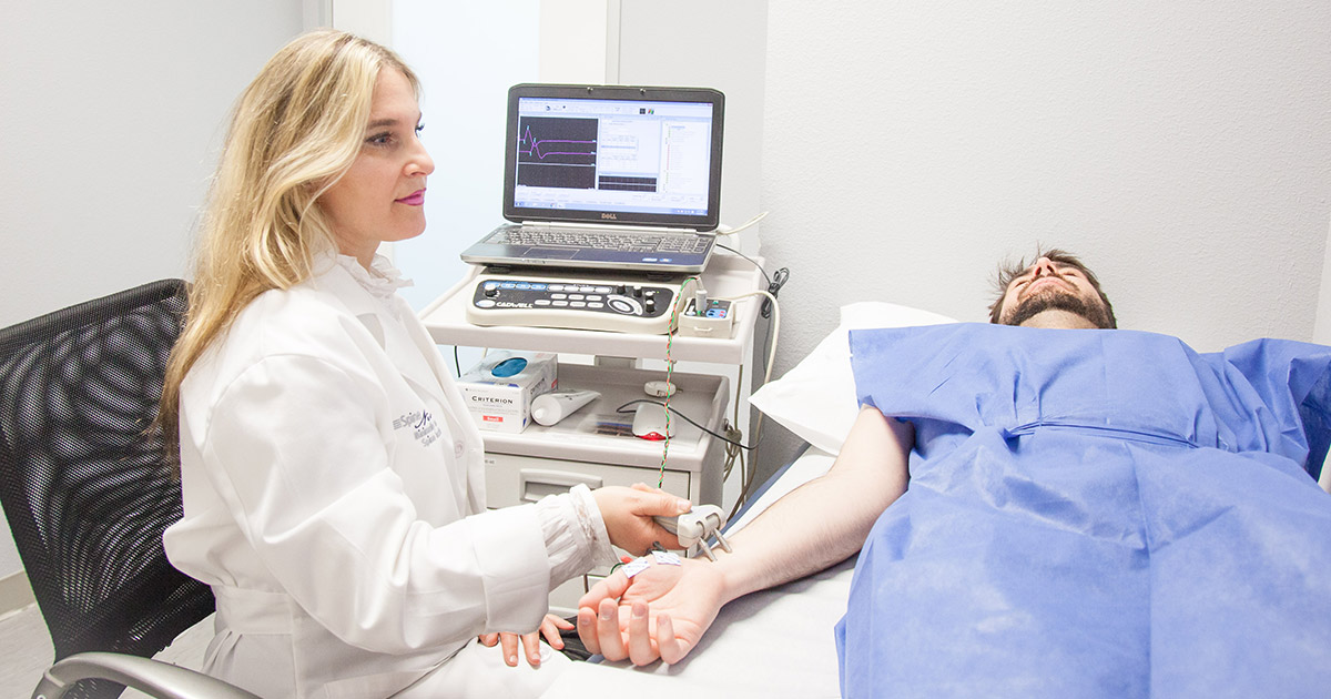
Schwannomas can be diagnosed using an electromyogram (EMG), or a test that measures the electrical impulses and activity of the muscles and nerves. This is a helpful tool because the electrical activity between nerves and muscles will be impaired when a schwannoma develops. This test measures the electrical activity when the muscles are in a forceful contraction, slight contraction, and at rest. A small needle with an electrode on the end is inserted into various muscles to perform an electromyogram. The individual having the electromyogram done will be asked to move the muscles being tested in various ways after the electrode has been placed. If an electromyogram is before a diagnostic imaging scan like an MRI or CT scan, the results may or may not prompt the use of one to pinpoint the tumor location. In addition, the results of a patient's electromyogram will determine if a biopsy on the area of concern should be performed. An electromyogram can also be used for a physician to rule out other diseases as the cause of symptoms an individual may be experiencing.
Get familiar with more ways to diagnose and treat schwannomas now.
Computerized Tomography Scan
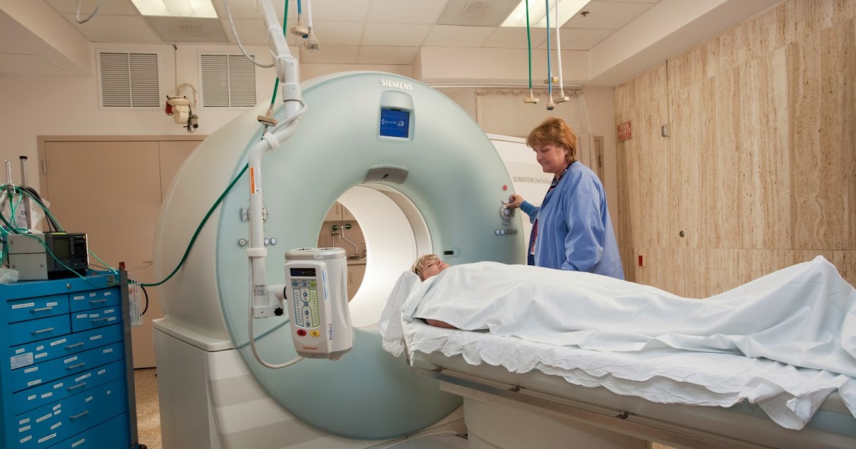
A computerized tomography scan (CT scan) will often be ordered when a physician suspects a patient may have a schwannoma. A CT scan utilizes special x-ray equipment in a procedure used to develop detailed scans or images of the targeted areas within the body. The images are formed when the CT machine takes continuous pictures in a helical fashion. Each picture developed during one of these scans will show the bones, organs, and other tissues within an individuals body. CT scans are utilized to check for thousands of possible abnormalities, one of them being the detection of abnormal growths like schwannomas. These scans are also used to determine what type of tissue damage has occurred around a schwannoma. This type of imaging test is extremely helpful for the planning of surgical procedures used to treat a schwannoma. With the administration of a CT scan, a physician can easily rule out other diseases or conditions that could be causing the same symptoms as a schwannoma.
Continue reading to learn about the methods of treating a schwannoma now.
Stereotactic Radiosurgery

Stereotactic radiosurgery is a type of procedure used in some cases to treat schwannomas. This type of radiosurgery is a precise type of therapeutic radiation used for the treatment of abnormalities in the central and peripheral nervous systems. Although the word surgery is included in the name of the procedure, no incision is needed for this kind of radiosurgery. This procedure works by targeting a specific area with powerful X-ray beams to shrink the abnormal tissues. The energy beams do this by destroying the DNA of the abnormal cells. This stops those cells from being able to grow or reproduce. This procedure is much less invasive than a traditional surgical excision, and it results in less damage to the surrounding tissues. This form of radiosurgery also has a lower risk for a patient to experience adverse side effects in comparison to traditional surgical excision of a schwannoma. When this procedure is used to treat a schwannoma, it is typically completed in one session. Radiosurgery to treat a schwannoma is most often used in individuals with schwannomas that affect the inner ear.
Get the details on more treatment options for schwannomas now.
Cancer Treatment
When a patient's schwannoma biopsy comes back as malignant or cancerous, the treatment will be geared in the same general direction as the treatment for other cancerous tumors. There are of a number of ways in which cancer treatment can be applied. The first priority of treating a cancerous schwannoma is to remove it surgically. If the location of the tumor or the surrounding tissues makes surgical excision exceptionally challenging, then radiation therapy will first be applied to shrink the tumor. After the tumor has been shrunk with radiation and removed, some patients may need subsequent chemotherapy. Chemotherapy is a classification of potent drugs that target and kill any cells in the body that are in the process of multiplying. This means even healthy noncancerous cells in the process of reproduction will be killed by chemotherapy in addition to the cancerous cells. Some patients with cancerous schwannomas may only require surgery to remove the tumor, or they may only need radiation therapy and surgery.
Learn more about treating schwannomas effectively now.
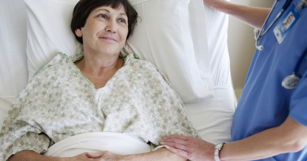
Physical Rehabilitation

Rehabilitation is a form of care that can help a patient regain, keep, or enhance abilities they need for everyday life. In the sense of physical rehabilitation, this consists of anything physical an individual does on a regular basis to meet their needs. Schwannomas can cause patients to be in chronic pain, lose their ability to mobilize, and or impair a certain part of their body. In addition, the treatment for schwannomas can also have side effects that cause a patient to lose their physical ability to perform everyday functions. In regions where treatment of a schwannoma results in the complete removal of one or more nerves, the patient may have to relearn how to use that particular muscle or set of muscles. The rehabilitation for issues caused by a schwannoma will have different goals and focuses from patient to patient. The main objective of this type of treatment is for the individual to regain their independence and normalcy in living their everyday life.
