How To Spot And Diagnose X-Linked Retinoschisis
X-linked retinoschisis is a genetically inherited condition that primarily affects individuals with XY chromosomes. The condition is most often diagnosed during childhood, sometimes when infants are as young as three months old. The most characteristic symptom is a reduction in vision that isn't improved with glasses. Some patients experience progressive vision loss over the course of their life, but others have fairly stable vision. X-linked retinoschisis occurs when a mutation in the RS1 gene located on the X chromosome causes the body to improperly produce retinoschisin. Normal retinoschisin is important to help the retina develop and be maintained throughout an individual's life. With X-linked retinoschisis, the retina's layers split, which disrupts the inter-cell communication and leads to loss of vision.
Read about the ways to spot and methods of diagnosing X-linked retinoschisis now.
Strabismus

Strabismus occurs when the eyes don't align properly. Normally, the two eyes work together to gather and interpret visual data. With strabismus, only one eye looks at the object the individual is viewing, while the other is aligned inward, upward, outward, or downward. Patients with X-linked retinoschisis may present with strabismus because of the separation of the layers of the retina. There are different types of strabismus. It can be intermittent or constant. Sometimes it affects only one eye, while other patients may alternate in the eye that focuses. To stop from developing double vision, the brain disregards the visual data that the misaligned eye provides, which tends to lead to 'lazy eye.'
Uncover more ways to spot X-linked retinoschisis now.
Farsightedness

Farsightedness, also called hyperopia, is a common vision-related condition. Some individuals with X-linked retinoschisis might have farsightedness that doesn't seem to get better with glasses. A farsighted individual sees objects in the distance clearly, but objects nearby tend to be blurry. Different degrees of farsightedness have an effect on the eye's ability to focus. When an individual has severe farsightedness, they might only be able to see objects that are far away, but those with mild farsightedness may see closer objects clearly. The structures in the eye that help focus objects are the cornea and lens. In normally-shaped eyes, these structures work together to focus images on the retina. However, in X-linked retinoschisis patients, the separation of the retinal layers causes images to be focused incorrectly.
Uncover more details on how to spot X-linked retinoschisis now.
Decline In Vision

One of the first symptoms of X-linked retinoschisis is a lack of visual acuity that can't be corrected with glasses. In cases where the condition is progressive, patients may experience a decline in vision that leads to legal blindness. When the retinal layers become separated, cystic macular lesions form between them. These are similar to blisters. The lesions might increase the thickness of the retina, which will make an individual's vision loss worse. However, they can be treated with different medications. Since the condition affects the retina's ability to develop, symptoms tend to present early in childhood. Children should have vision tests at six months old, three years old, prior to first grade, and every two years throughout their school years.
Learn about how X-linked retinoschisis is diagnosed now.
Electroretinography
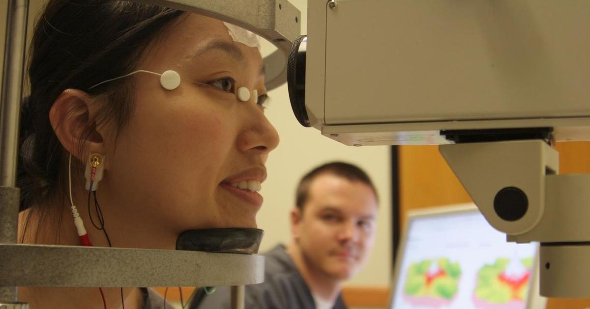
Electroretinography is a diagnostic eye test that determines how well the retina functions. The retina has multiple layers of specialized cells. The rods and cones are photoreceptor cells that detect light. There are also ganglion cells to transmit visual data to the brain. Electroretinography detects electrical signals given off by the photoreceptors, plus electrical signals from the bipolar and Muller cells located between the ganglion and photoreceptor cells. When the readings are abnormal, they can help an ophthalmologist determine what retinal abnormalities are causing vision loss. The test is done by placing an electrode on a patient's cornea to measure the eye's electrical responses to light.
Keep reading for more information on how X-linked retinoschisis is diagnosed now.
Visual Field Test
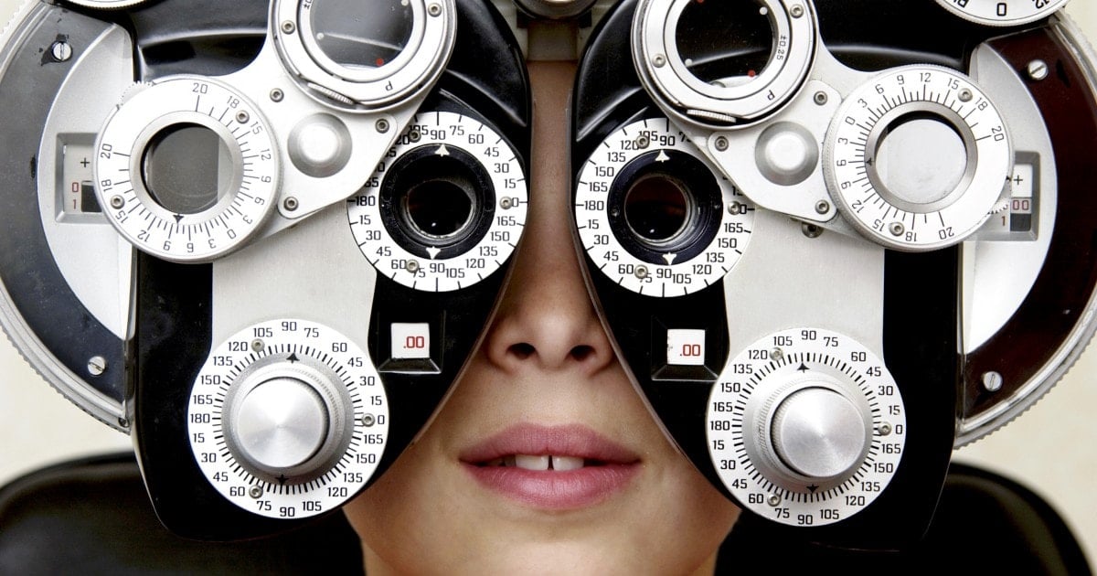
A visual field test is used to determine whether an individual has blind spots in their vision and where the blind spots are located. The visual field is the amount of area an eye can see when an individual focuses on one central point. Visual field testing is one of the ways an ophthalmologist measures the amount of vision an individual has in either of their eyes, and the amount of vision loss that might have occurred. A confrontational visual field test is done by having a patient cover one eye and look at a central point with another. The doctor will hold up different numbers of fingers in the patient's peripheral vision to determine whether they can see them. An automated perimetry test might be used to gauge how well individuals see objects in their field of vision. This type of test creates more detailed maps of an individual's blind spots.
