Guide To The Structure Of The Heart

August 27, 2024

Located in the upper portion of the chest, the heart sits behind the breastbone and is responsible for pumping blood throughout the entire body. A healthy heart is roughly the size of a fist, and it usually beats between sixty to one hundred beats each minute. Over twenty-four hours, the heart circulates up to two thousand gallons of blood around the body. The typical weight for the adult heart is between seven to fifteen ounces. To evaluate a patient's heart health, doctors often begin by measuring the patient's heart rate. The patient will also have their blood pressure recorded, and the clinician might use a stethoscope to check for any abnormal heart sounds or rhythms. To get more detailed information, patients may have an electrocardiogram (a recording of the heart's electrical activity), and an echocardiogram (an ultrasound of the heart) may be recommended to learn more about how well the patient's heart is functioning. Patients with heart problems are typically treated by cardiologists, physicians who specialize in cardiac care.

Atria
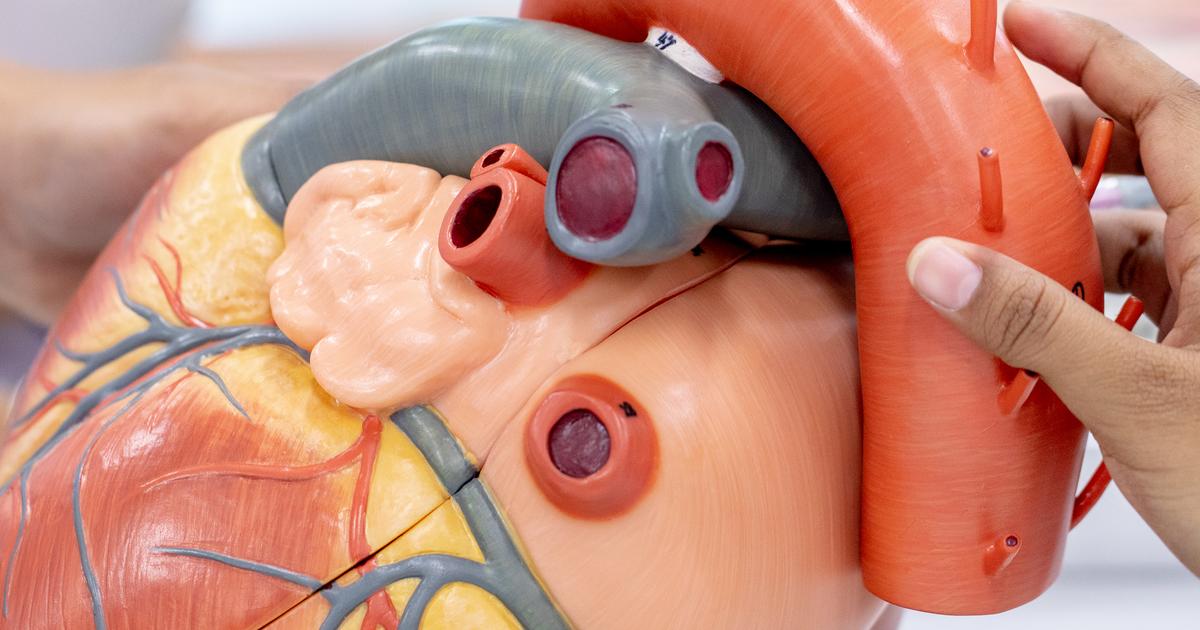
Photo Credit: Dreamstime

The atria are the upper left and right chambers of the heart. Separated by a structure known as the interatrial septum, these two chambers work together to receive blood as it returns to the heart from other areas of the body. Specifically, the right atrium receives de-oxygenated blood returning from the head, neck, arms, and chest via a major vessel known as the superior vena cava. De-oxygenated blood from the lower body, including the legs, back, abdomen, and pelvis, is returned to the right atrium through the inferior vena cava. The left atrium receives blood that returns to the heart from the pulmonary veins. These veins connect the left atrium to the lungs. Unlike the blood that returns to the right atrium, this blood is full of oxygen. The right atrium contains nodes that generate the electrical impulses that regulate the rate and rhythm of the heart. Known as the heart's pacemaker, the sinoatrial node is located in the upper wall of the right atrium. The electrical impulses that travel from this location eventually reach the atrioventricular node in the lower part of the right atrium. The atrioventricular node delays the impulses from the sinoatrial node by a fraction of a second. During this time, the atria contract, and blood is sent to the ventricles. Atrial fibrillation and atrial flutter are two conditions that can affect the atria.

Ventricles

Photo Credit: Dreamstime

The left and right ventricles compose the lower portion of the heart, and these chambers are responsible for pumping blood to the body. Blood from the right atrium passes into the right ventricle through the tricuspid valve. After the blood arrives in the right ventricle, contractions of the ventricles open the pulmonary valve, and blood is pumped into the main pulmonary artery. The heart's mitral valve transports blood from the left atrium to the left ventricle. When the ventricles contract, blood in the left ventricle enters the aortic valve and is pumped directly to the aorta. Oxygen-rich blood is carried by the aorta to the rest of the body. The walls of the ventricles are thicker than those of the atria because they need more power for contraction. The electrical impulses sent by the nodes in the atria are relayed to the ventricles through tissues known as Purkinje fibers, and this stimulates contraction. Ventricular fibrillation, ventricular tachycardia, and heart failure are some of the cardiac conditions that impact the ventricles.

Heart Wall

Photo Credit: Dreamstime

The heart wall has three layers. In addition to protecting the heart, the heart wall coordinates synchronization of the heartbeat and enables the heart to contract. The wall is composed of cardiac muscle, connective tissue, and endothelium. Known as the epicardium or visceral pericardium, the outer layer of the heart wall forms the inner layer of the pericardium, a sac that surrounds and protects the heart. The majority of the epicardium is formed of connective tissues, including fat. Coronary blood vessels located in the epicardium supply the heart wall with blood, and the epicardium also helps in the production of pericardial fluid. The myocardium is the middle layer of the heart wall, and it is also the thickest layer. This part of the heart wall is constructed of cardiac muscle that stimulates heart contractions. The endocardium is the innermost layer of the heart wall, and it is the thinnest of all the layers. It serves as a protective covering for the valves of the heart, and also acts as a lining for the heart chambers. Endocarditis, an infection of the endocardium, is one of the major issues that could arise in this area of the heart.

Arteries And Veins

Photo Credit: Dreamstime

Blood vessels (arteries and veins) are hollow tubes that carry blood throughout the body. Many of the major arteries in the body branch off from the aorta, the body's largest artery. This artery runs from the top of the left ventricle to the abdomen, and it is roughly twelve inches long and one inch in diameter. Branching off the aorta, the brachiocephalic artery carries oxygenated blood from the aorta to the head, neck, and arms. The carotid arteries also supply oxygenated blood to the head and neck, and the subclavian arteries provide oxygenated blood to the arms. The coronary arteries transport oxygenated blood to the heart muscle, and the common iliac arteries carry oxygenated blood from the abdominal part of the aorta to the legs and feet. The pulmonary artery carries deoxygenated blood to the lungs from the right ventricle of the heart. The brachiocephalic veins are two major veins that join together to create the superior vena cava, and the common iliac veins conjoin to form the inferior vena cava. Together, the venae cavae work to transport deoxygenated blood from many areas of the body back to the heart. The pulmonary veins are responsible for transporting oxygenated blood from the lungs directly to the heart.

Electrical System

Photo Credit: Dreamstime

The electrical system of the heart triggers and regulates the heartbeat in a process known as cardiac conduction. Factors such as exercise, temperature, and hormones influence this process. In the first step of cardiac conduction, nerve impulses are generated by the sinoatrial node. They travel through the entire heart wall, causing both of the atria to contract. In the second stage of conduction, the nerve impulses reach the atrioventricular node and are delayed for roughly one-tenth of a second. This delay triggers atrial contraction, and blood from the atria is emptied into the ventricles. Next, the nerve impulses travel from the atrioventricular node to the atrioventricular bundle, and they are carried to the left and right ventricles and the center of the heart. As the impulses travel down the atrioventricular bundle, they eventually reach the Purkinje fibers and trigger the contraction of the ventricles. Cardiac conduction controls the cardiac cycle, the sequence of events that occurs when the heart beats. In the diastole phase of the cardiac cycle, the atria and ventricles are relaxed, and blood flows into them. During the systole phase, the ventricles contract, allowing blood to be sent from the heart to the rest of the body.

MORE FROM HealthPrep