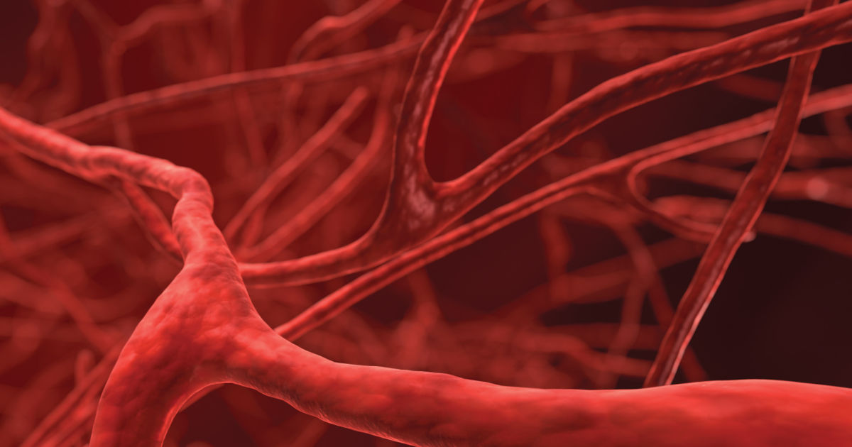
What Are The Types Of Veins?
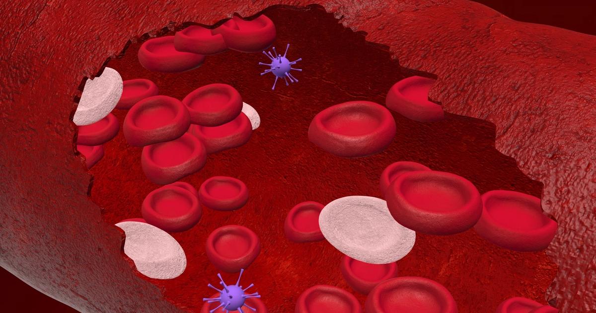
A vein is a vessel structure that exists throughout the body that transports blood from all parts of the body to the heart. Arterial blood moves through a high-pressure mechanism away from the heart, and the venous blood moves through a low-pressure mechanism and muscle contractions back to the heart. Vein diameter ranges from one millimeter to one and a half centimeters. Veins contain three tissue layers in their walls. Connective tissue, elastic fibers, and collagen fibers make up the tunica adventitia or strong outer layer of the veins and arteries. Elastic fibers and smooth muscle form the tunica media or middle layer of veins and arteries. Smooth endothelium, elastic tissues, and an elastic membrane lining form the tunica intima or innermost layer of veins and arteries. Veins contain muscular structures in the tunica intima that move blood against gravity back to the heart. Veins are classified according to their location and size.
Pulmonary Veins
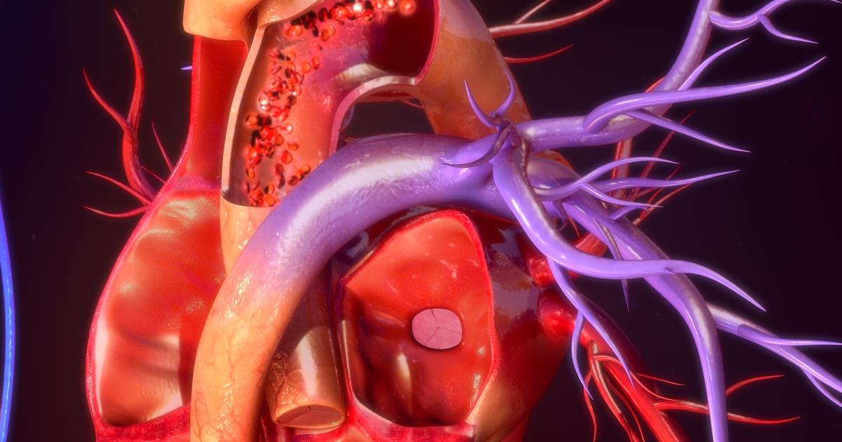
Pulmonary veins are a type of large-sized vein in the body that serves as the channel where newly oxygenated blood from the lungs moves into the heart before being pumped around to the rest of the body. The pulmonary veins are the only veins in an individual's body that transport oxygenated blood. While an individual is still in the womb, their blood flow bypasses the pulmonary veins because their lungs are not exposed to air yet. Once the individual is born, the pulmonary veins open up, and blood is then routed through the lungs and back to the heart through the pulmonary veins. All veins in an individual's body carry blood to the heart regardless of its oxygen concentration, which is why pulmonary veins are considered veins even though they transport oxygenated blood. The pulmonary veins also exhibit more structural characteristics of a vein rather than those of an artery. The four pulmonary veins are connected to both of an individual's lungs and run to the left atria of the heart. From the left atria, the oxygenated blood moves to the left ventricle and is pumped out of the heart into the aorta. The pulmonary veins are typically around one centimeter in diameter, but they may be smaller in females.
Systemic Veins
Systemic veins are the veins that make up an individual's systemic circuit that circulates the body and then carries blood to the pulmonary circuit that starts with entry into the right atrium of the heart. An individual's systemic circuit begins when blood leaves their heart through the aorta. Systemic arteries are vessels that carry blood from the aorta to all the different body tissues, and the systemic veins are the vessels that take the oxygen-poor blood back to the heart. The systemic veins begin in the smallest veins, which are referred to as superficial veins, where oxygen-poor blood moves away from the most distant tissues in the direction of larger sized veins, which are referred to as the connecting veins. The oxygen-poor blood moves from the connecting veins into the deep veins, which eventually merge into the main systemic veins. The two main systemic veins are called the inferior vena cava and the superior vena cava. Both the inferior vena cava and superior vena cava move the oxygen-poor blood from the deep veins and up into the right atrium of the heart, ending the systemic venous circuit.
Superficial Veins
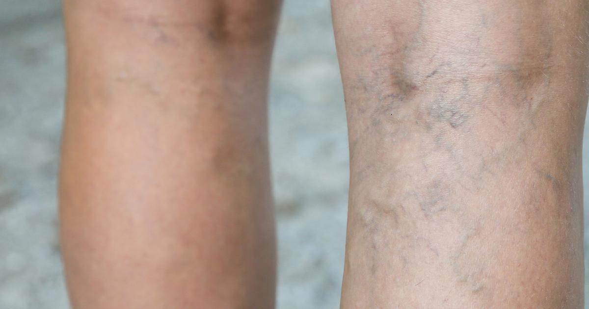
Superficial veins are a type of smaller vein in the body. They are distant from corresponding arteries and closer to the skin surface. Superficial veins differ from the deep veins because they are not directly paired with any of the arteries nearby. The superficial veins are typically located in the fatty layer that sits under the skin. The superficial veins not only deliver oxygen and nutrients to the skin tissues and the tissues underneath it, but they also have a critical physiological function. When an individual's body becomes too hot, blood is forced from their deep veins to the superficial veins to expedite the process of heat transfer from the blood to the skin in the form of sweat. Superficial veins can often be seen under the skin as blue or green in fair-skinned individuals and dark red or purple in olive skin tones. Blood moves slower in the superficial veins because even though they contain one-way valves, they are not surrounded by muscular tissue that helps push the blood at a faster rate the same way deep veins are.
Deep Veins
Deep veins vary in size and supply deep layers of bone and muscle tissue close to a corresponding artery. Deep veins are found far under the skin going through the muscles and running alongside an individual's bones. Blood in the deep veins can move faster and more efficiently than it can in superficial veins because the deep veins are surrounded by muscular tissue that helps the valves squeeze blood toward the heart. The function of the valves inside of the deep veins and the muscular tissues around them is critical because gravity is a constant force against their direction of motion. Deep veins can hold more blood at any given time than the arteries because the venous walls are thinner and more flexible than the arterial walls. Some of the deep veins present in an individual's lower extremities are the internal iliac, pelvic vein, deep femoral vein, external iliac, common iliac, and the popliteal veins. Some deep veins found in the upper extremities include the ulnar vein, axillary vein, radial vein, brachial vein, and subclavian veins.
Connecting Veins
Connecting or communicating veins are short veins that carry blood from the superficial veins near the skin to the deep veins in the muscles and organs. Communicating veins are also known to connect one deep vein to another deep vein in the body. Connecting veins can be classified or divided into two subgroups. Communicating veins are connecting veins that run from superficial veins or a deep vein to another deep vein when they are located within the same fascia compartment of the body. Perforating veins are connecting veins that link superficial veins or deep vein to another deep vein located in a different fascia compartment of the body. To facilitate this action between compartments in the body, there are numerous holes the perforating connecting veins flow through to reach their destination. Connecting veins also contain small valves to keep the blood moving in one direction toward the larger veins that flow to the heart.
