Guide To The Structure Of The Heart
Heart Wall

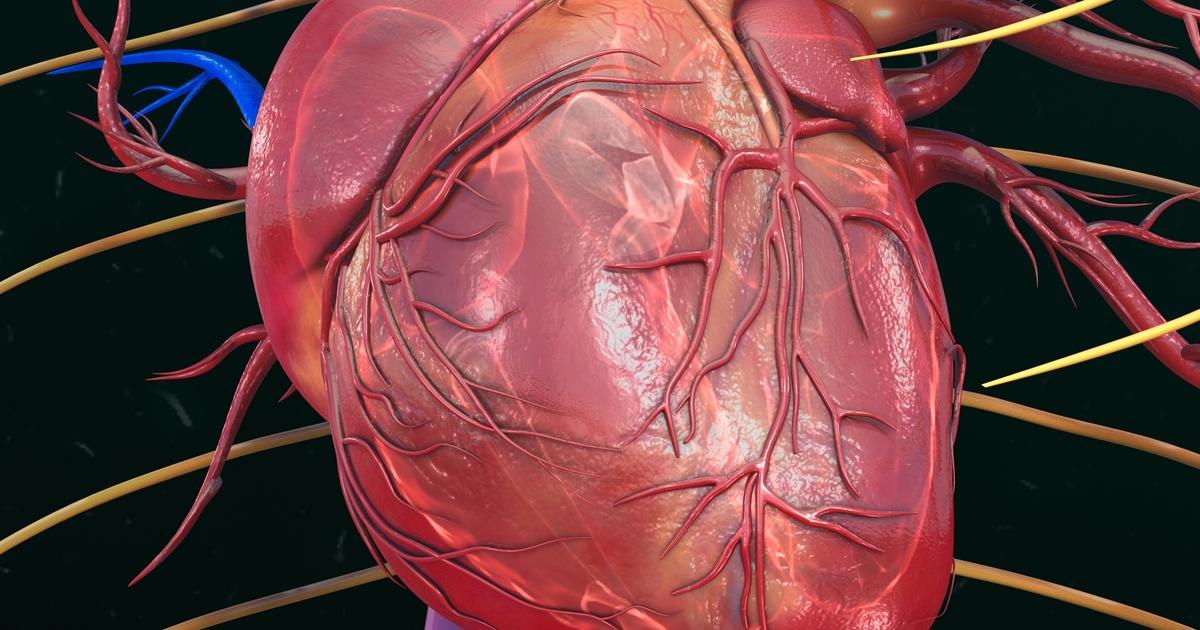
The heart wall has three layers. In addition to protecting the heart, the heart wall coordinates synchronization of the heartbeat and enables the heart to contract. The wall is composed of cardiac muscle, connective tissue, and endothelium. Known as the epicardium or visceral pericardium, the outer layer of the heart wall forms the inner layer of the pericardium, a sac that surrounds and protects the heart. The majority of the epicardium is formed of connective tissues, including fat. Coronary blood vessels located in the epicardium supply the heart wall with blood, and the epicardium also helps in the production of pericardial fluid. The myocardium is the middle layer of the heart wall, and it is also the thickest layer. This part of the heart wall is constructed of cardiac muscle that stimulates heart contractions. The endocardium is the innermost layer of the heart wall, and it is the thinnest of all the layers. It serves as a protective covering for the valves of the heart, and also acts as a lining for the heart chambers. Endocarditis, an infection of the endocardium, is one of the major issues that could arise in this area of the heart.
Arteries And Veins

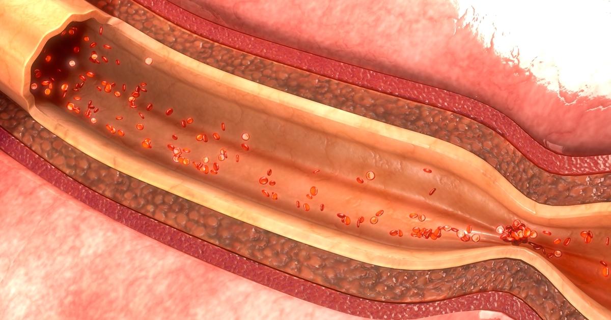
Blood vessels (arteries and veins) are hollow tubes that carry blood throughout the body. Many of the major arteries in the body branch off from the aorta, the body's largest artery. This artery runs from the top of the left ventricle to the abdomen, and it is roughly twelve inches long and one inch in diameter. Branching off the aorta, the brachiocephalic artery carries oxygenated blood from the aorta to the head, neck, and arms. The carotid arteries also supply oxygenated blood to the head and neck, and the subclavian arteries provide oxygenated blood to the arms. The coronary arteries transport oxygenated blood to the heart muscle, and the common iliac arteries carry oxygenated blood from the abdominal part of the aorta to the legs and feet. The pulmonary artery carries deoxygenated blood to the lungs from the right ventricle of the heart. The brachiocephalic veins are two major veins that join together to create the superior vena cava, and the common iliac veins conjoin to form the inferior vena cava. Together, the venae cavae work to transport deoxygenated blood from many areas of the body back to the heart. The pulmonary veins are responsible for transporting oxygenated blood from the lungs directly to the heart.
