Watch Out For These Early Warning Signs Of Glaucoma
Patchy Blind Spots
An individual who begins to experience the appearance of patchy blind spots may be affected by glaucoma. These patchy blind spots typically occur when an individual's glaucoma has progressed to the degree of causing their optic nerve to incur damage. The optic nerve is the component of the eye responsible for the transmission of visual information to the vision centers in the brain. The optic nerve contains over a million tiny nerve fibers that originate from the retina. The fluid an individual's eye produces (aqueous humor) is meant to drain from the eye through a net-like structure, and it then flows into the individual's blood stream. Most individuals affected by glaucoma have a problem with this system of aqueous humor drainage from the eye. The aqueous humor is unable to drain and begins to accumulate in the eye, compressing the small fibers of the optic nerve and other components of the eye. Over time, this malfunction leads to permanent damage as the optic nerve fibers begin to die off. This mechanism is what causes a glaucoma patient to see patchy blind spots in their peripheral vision.
Mid-Dilated Pupil

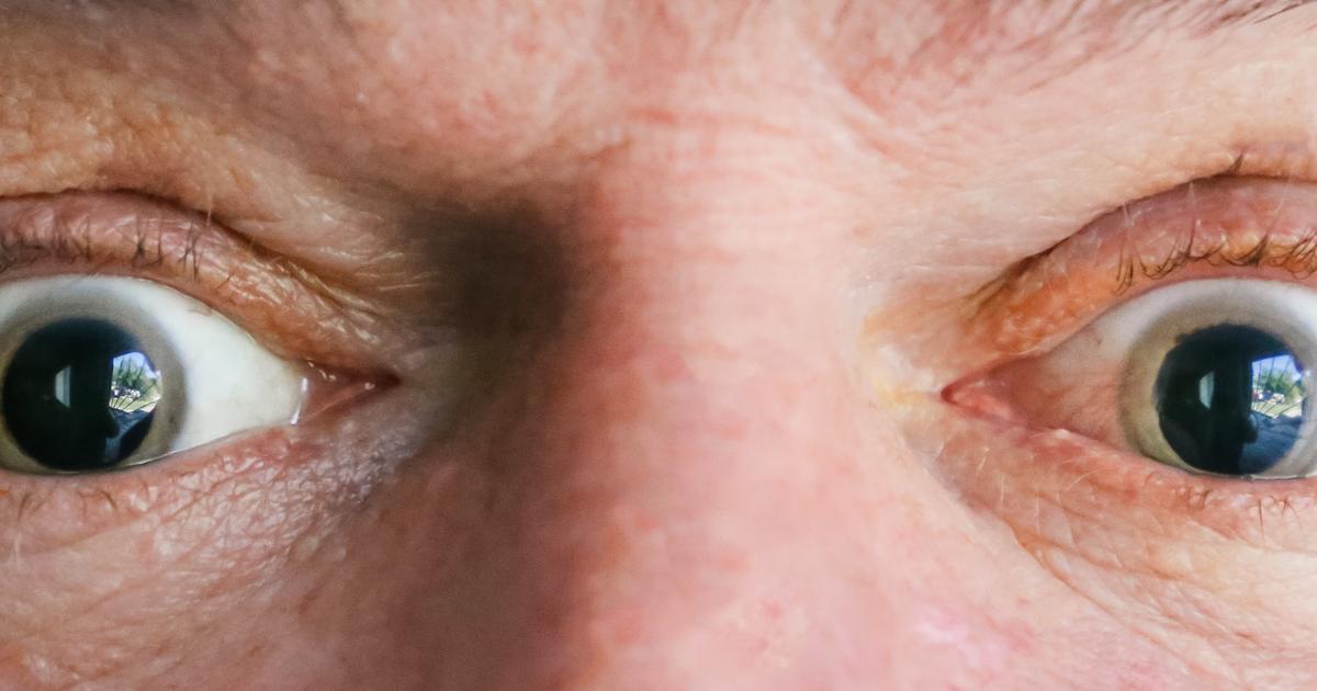
A mid-dilated pupil in an individual's eye can indicate they are suffering from angle-closure glaucoma. This type of glaucoma occurs when the eye cannot drain aqueous humor due to a specific mechanism. The iris in the eye is the part that contains color. The pupil is the black structure in the center of the iris. The lens of the eye sits right behind the pupil and iris and is held in place by ligaments. The ciliary body is the muscular structure that sits behind the iris that produces aqueous humor, which flows into the cornea and pupil and is then drained through the net-like structure at the junction where the iris and cornea meet. The mid-dilated pupil happens during a sudden occurrence of angle-closure because most contact between the lens and the iris happens when an individual's pupil is in a mid-dilated position. The net-like structure where the aqueous humor should drain from becomes obstructed, and the pressure in the eye increases. This increasing pressure causes a restriction of blood supply to the muscle that controls the dilation of the pupil. The muscle becomes non-functional as a result, and the pupil remains in the mid-dilated position. This mechanism is why a mid-dilated pupil can indicate acute angle closure glaucoma.
