Guide To Positron Emission Tomography Scans (PET Scans)

January 31, 2023

A positron emission tomography scan (PET scan) is a type of imaging test used for diagnosing numerous diseases in an individual's body. This scan highlights parts of the body where there are increased rates of certain chemical activities. A PET scan can tell a patient's doctor about how their body uses oxygen, how their body processes glucose, and about their blood flow. These scans can give a patient's doctor insight into problems occurring at the cellular level. This helps them identify and evaluate certain complex systemic conditions and diseases, including heart problems and brain disorders. Around two million PET scans are performed annually in the United States.

Individuals need these scans. As they diagnose many conditions, they are vital for receiving the necessary treatment. For instance, these scans help direct the best cancer treatment, including radiation therapy, chemotherapy, and immunotherapy for cancer. The scans show the effectiveness of the treatment as well. These scans also accurately diagnose heart conditions, helping doctors determine where surgery and medications for heart disease are appropriate. Of course, patients must understand how positron emission tomography scans work first.

How The Scan Works

A positron emission tomography scan uses a large machine with a hole in the center or a scanning device. Both will pick up subatomic particles or photons emitted by a radiotracer in the tissues or organ being examined. The radiotracer used for this scan depends on the particular tissues or organs of interest and the scan's purpose. The selected radiotracer is administered to the patient's body through a vein in their arm via an intravenous line. Once the radiotracer has been administered, the scanning part of the device slowly moves over the necessary part of the patient's body.

As particular tissues in the body break down the radiotracer, positrons are emitted. When positrons are emitted in the body from the breakdown of the radiotracer, gamma rays are produced. The positron emission tomography scanner can pick up these gamma rays and use this information to compose an image map of the internal tissues. The higher the concentration of gamma rays, the brighter the spot will appear in the scan's image.

Types Of PET Scans

Many individuals will receive a standard positron emission tomography scan. Of course, the precise part of the patient's body that doctors want to examine may change. However, there are different types of PET scans that are often considered to have special significance. One of these scans is the cardiac positron emission tomography scan, which is also referred to as a SPECT scan. It is a cardiac molecular imaging scan. In this type, the doctor will inject patients with a radioactive tracer that will circulate to their heart. Then, the patient will receive a scan of their heart. This scan detects the tracer's signal. The result is a three-dimensional image.

Many positron emission tomography scans use an injection of fludeoxyglucose (FDG) as the radioactive tracer. This injection is of particular prominence in PET f-18 FDG scans. This scan involves a glucose scan immediately following the positron emission tomography scan. The reason that this is done, according to reports, is to determine whether or not there is permanent heart muscle damage. In addition to these types, it is worth noting that there is also a PET/CT scan. This scan combines a positron emission tomography scan with a computerized tomography scan. Doctors use this more a more accurate diagnosis.

When PET Scans Are Used

Positron emission tomography (PET) scans are utilized to diagnose and evaluate the progress of numerous different disorders, diseases, and conditions. The most common use for these scans is for the detection, diagnosis, and progression of cancer in the body and evaluation of cancer treatments. Another reason this type of scan may be performed is to diagnose conditions associated with the deterioration of brain function. Examples include Parkinson's disease, Huntington's disease, Alzheimer's disease, epilepsy, cerebrovascular accidents, and other types of dementia.

This type of scan can also be used to evaluate the flow of blood to the muscle tissues of a patient's heart to help evaluate the efficacy of a therapeutic procedure to help blood flow to the heart muscle. Positron emission tomography scans can also help further identify masses or lesions seen in a patient's lung tissues picked up on a chest CT or chest x-ray.

Preparing For A PET Scan

This type of scan is done on an outpatient basis, meaning the patient who undergoes the test will be able to go home immediately afterward. Before having a positron emission tomography scan, a patient should inform their physician of all vitamins, herbal remedies, supplements, and medications they are taking. Some medications, supplements, and vitamins may require patients to follow extra instructions before their scan.

Patients should let their doctor known if they are pregnant or if there is a possibility they are pregnant. In most cases, patients will be instructed not to consume any food for at least six hours before their scan. If the positron emission tomography scan involves any part of their heart, they are usually advised to avoid consuming caffeine for twenty-four hours before their scan.

Recovering From The Scan

After a positron emission tomography scan, patients can often resume their normal activities. This only changes if the patient's doctor gives them alternative instructions. Despite being able to resume normal activities, there are still some instructions to help patients recover from a positron emission tomography scan. The radioactive tracer used for the scan will stay in the patient's body for an extended period afterward. Patients need to drink a significant amount of fluids to help flush the tracer from their system. In most cases, they can do so in a couple of days, perhaps less. Besides this, most patients do not have additional instructions for recovery. They will often receive their results in a few days at a follow-up appointment with their doctor.

Compared To CT And MRI Scans
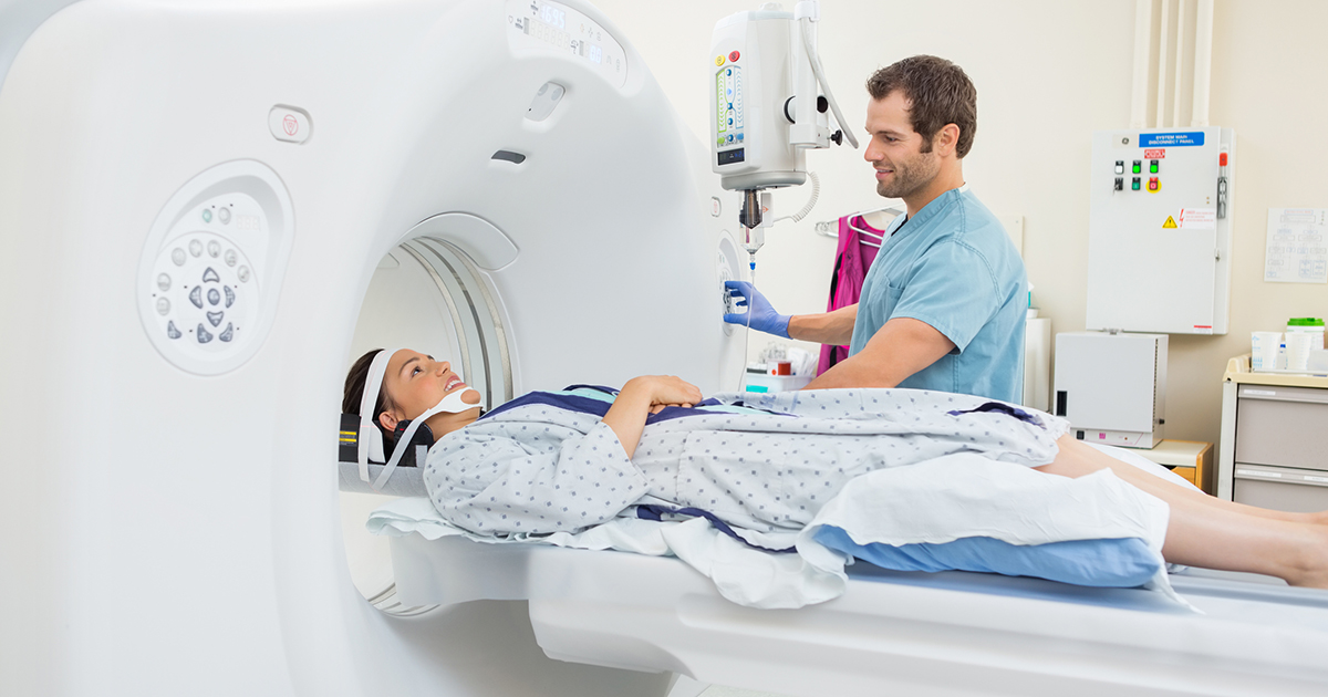
Positron emission tomography scans can reveal problems occurring at the cellular level of a patient's body, unlike magnetic resonance imaging (MRI) and computerized tomography (CT) scans. Computerized tomography scans utilize special x-ray equipment to compose detailed images of the inside of a patient's body. Magnetic resonance imaging uses radiofrequency pulses and magnetic fields to produce detailed images of the patient's internal structures like bone, organs, and other soft tissues.

CT scans and MRI scans can only reveal changes in a patient's body that occur at later stages of their respective disorder or disease. These scans can pick up changes in a patient's body once the disease or condition begins to alter the structure of the tissues or organs in their body. In some cases, an individual must have PET-MRI scans or PET-CT scans. Combining an MRI or CT scan with a positron emission tomography scan allows a computer to mesh the images for a more accurate diagnosis.

Risks Of A PET Scan

Positron emission tomography scans have fewer risks associated with them than those of MRI scans, CT scans, and standard x-rays. A minimal amount of radiation exposure will occur during a positron emission tomography scan because a radiotracer chemical is used. Radiotracer chemicals may also be referred to as radionuclides, radiopharmaceuticals, or radioactive tracers. Radiotracers used in positron emission tomography scans are a molecule that contains glucose and an attached radioactive component.

A patient can have an allergic reaction to the substances used as the radioactive tracer. Individuals who are allergic to aspartame, saccharin, and iodine are at a greater risk of experiencing problems during their positron emission tomography scan. Asthma, heart disease, dehydration, and kidney disease patients at a greater risk of experiencing complications from a PET scan. This also applies to those who take beta-blockers, nonsteroidal anti-inflammatories, or interleukin-2.

PET Scans And Pregnancy

In the vast majority of cases, the benefits of positron emission tomography scans outweigh the risks associated with them. However, this may not be the case for many pregnant women. Many doctors say that the radioactive material used in these scans can negatively affect the unborn baby. Thus, pregnant women often do not undergo positron emission tomography scans except in extremely specific circumstances.

The effect of the radioactive material on babies is also why women must take precautions when they are breastfeeding. In some cases, doctors will order a positron emission tomography scan for a woman who is breastfeeding. These women often need to pump and discard their breast milk until the radioactive material is gone. Women need to consult their doctor on the timing of this. Finally, some patients who have gotten a positron emission tomography scan will need to avoid pregnant women and small children for a few hours. Once again, they should ask their doctor about this.

PET Scans And Diabetes

Diabetes patients are still able to undergo positron emission tomography scans. However, these individuals will need to follow alternative instructions before their scans, and often afterward as well. This is because the normal instructions can interfere with the patient's blood sugar. Of course, as all individuals with diabetes know, managing their blood sugar is vital. Patients must consult their doctor before a positron emission tomography scan for instructions that are specific to their needs. There are, though, some common ones.

In most cases, diabetes patients will be asked to take their normal insulin dose. They will also consume a light meal four hours prior to their positron emission tomography scan. After the scan, diabetes patients typically follow the same procedure as other individuals. However, their doctor will tell them if they need to do anything different.

PET Scans And Claustrophobia

The machines used for positron emission tomography scans often trigger claustrophobia for patients. They are enclosed, which is not great for individuals with a fear of small spaces. The symptoms of claustrophobia during one of these scans include chills, sweating, excessive fear, nausea, dizziness, feeling faint, and an increased heart rate.

Thankfully, patients who are concerned about feeling claustrophobic during this scan can manage this reaction. Patients can choose to listen to music in certain cases, which allows them to focus on the lyrics instead of the scan. Deep breathing can help as well. In serious cases, some patients can receive mild sedative medication to keep themselves calm during the scan. Doctors will prescribe and administer this medication as needed.

MORE FROM HealthPrep