Guide To Positron Emission Tomography Scans (PET Scans)
A positron emission tomography scan (PET scan) is a type of imaging test used for diagnosing numerous diseases in an individual's body. This scan highlights parts of the body where there are increased rates of certain chemical activities. A PET scan can tell a patient's doctor about how their body uses oxygen, how their body processes glucose, and about their blood flow. These scans can give a patient's doctor insight into problems occurring at the cellular level. This helps them identify and evaluate certain complex systemic conditions and diseases, including heart problems and brain disorders. Around two million PET scans are performed annually in the United States.
Individuals need these scans. As they diagnose many conditions, they are vital for receiving the necessary treatment. For instance, these scans help direct the best cancer treatment, including radiation therapy, chemotherapy, and immunotherapy for cancer. The scans show the effectiveness of the treatment as well. These scans also accurately diagnose heart conditions, helping doctors determine where surgery and medications for heart disease are appropriate. Of course, patients must understand how positron emission tomography scans work first.
How The Scan Works

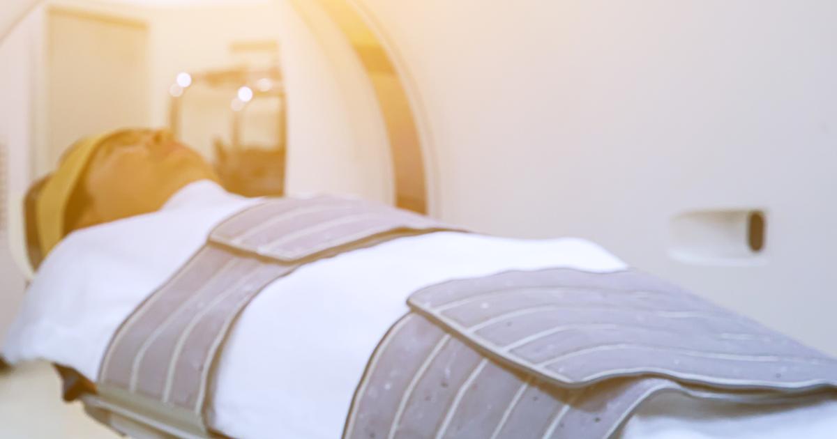
A positron emission tomography scan uses a large machine with a hole in the center or a scanning device. Both will pick up subatomic particles or photons emitted by a radiotracer in the tissues or organ being examined. The radiotracer used for this scan depends on the particular tissues or organs of interest and the scan's purpose. The selected radiotracer is administered to the patient's body through a vein in their arm via an intravenous line. Once the radiotracer has been administered, the scanning part of the device slowly moves over the necessary part of the patient's body.
As particular tissues in the body break down the radiotracer, positrons are emitted. When positrons are emitted in the body from the breakdown of the radiotracer, gamma rays are produced. The positron emission tomography scanner can pick up these gamma rays and use this information to compose an image map of the internal tissues. The higher the concentration of gamma rays, the brighter the spot will appear in the scan's image.
Types Of PET Scans

Many individuals will receive a standard positron emission tomography scan. Of course, the precise part of the patient's body that doctors want to examine may change. However, there are different types of PET scans that are often considered to have special significance. One of these scans is the cardiac positron emission tomography scan, which is also referred to as a SPECT scan. It is a cardiac molecular imaging scan. In this type, the doctor will inject patients with a radioactive tracer that will circulate to their heart. Then, the patient will receive a scan of their heart. This scan detects the tracer's signal. The result is a three-dimensional image.
Many positron emission tomography scans use an injection of fludeoxyglucose (FDG) as the radioactive tracer. This injection is of particular prominence in PET f-18 FDG scans. This scan involves a glucose scan immediately following the positron emission tomography scan. The reason that this is done, according to reports, is to determine whether or not there is permanent heart muscle damage. In addition to these types, it is worth noting that there is also a PET/CT scan. This scan combines a positron emission tomography scan with a computerized tomography scan. Doctors use this more a more accurate diagnosis.
