Causes And Complications Of Fibrous Dysplasia
Fibrous dysplasia is a disorder of the bone where an individual's healthy bone tissue is replaced with fibrous or scar-like tissue. As a result, a fibrous dysplasia patient will have weaker bones than other individuals. These weakened bones can experience fractures and or become deformed. It is more common for fibrous dysplasia to develop in one bone at one site, but it can develop in multiple bones or at multiple sites. The most common bones affected by fibrous dysplasia include the shinbones, thighbone, pelvis, skull, ribs, and upper arm bone. Symptoms of fibrous dysplasia include swelling, bone pain, leg bone curvature, arm fractures, leg fractures, and deformities of the bone. Fibrous dysplasia is diagnosed with the use of x-rays, bone scans, bone biopsy, CT scans, and an MRI.
Genetic Mutation
Fibrous dysplasia is thought to be caused by a specific gene mutation that spontaneously occurs following embryonic fertilization. This type of genetic mutation is not one inherited from the patient's parents. The mutated gene that causes the development of fibrous dysplasia is a gene called GNAS1. Part of the cells in an affected individual's body contains a healthy copy of this gene, while the rest of the cells carry the mutated copy of the gene. This mutation occurs in a gene responsible for the production of a particular protein, called the G-protein. The mutation causes the affected individual's body to produce an excessive amount of this G-protein. This protein is responsible for the production of a molecule called cyclic adenosine monophosphate, which is associated with the process the body uses to recycle bone tissue and the bone turnover that results from it. The bone is broken down properly in this process in affected individuals but is thought to be replaced by fibrous tissue rather than new bone tissue.
Vision Loss
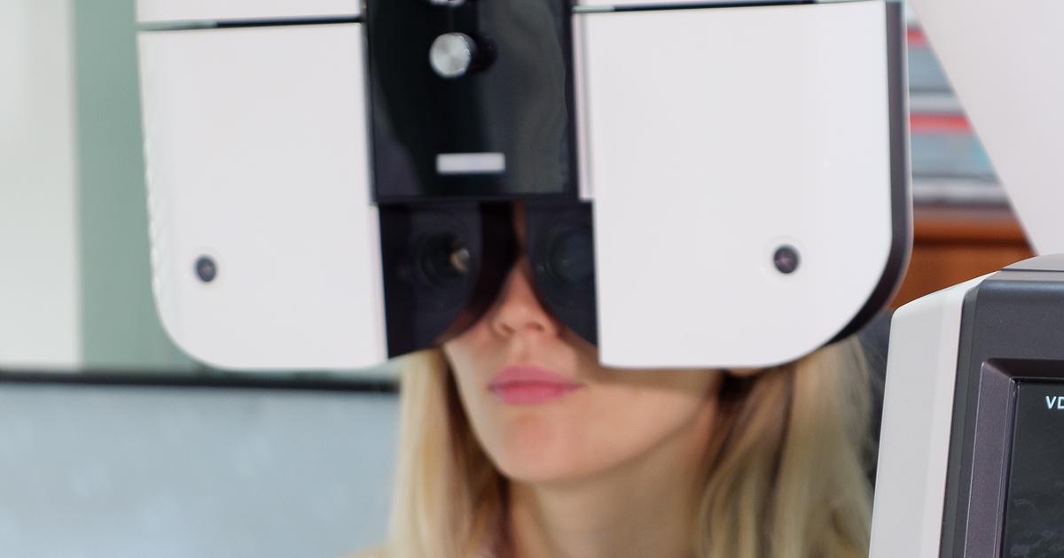
Individuals with fibrous dysplasia can experience vision loss, as the fibrous lesions that replace bone tissues can develop on any bone, including the craniofacial bones and skull. Lesions that develop in the facial bones near the individual's eyes can cause their eyes to become misaligned, making it difficult to see correctly. The optic nerve that connects an individual's eyes to the brain tissue can be affected when fibrous lesions grow nearby it. A patient's optic nerve can become compressed as a result of this malfunction. The optic nerve is responsible for transmitting signals that contain visual information to the vision center in the brain. It is uncommon for this mechanism to occur in both eyes, as separate parts of the same bones surround the eyeball and its corresponding structures. While general impairment of the vision and double vision are symptoms that occur more commonly, vision loss can occur in individuals who have severe cases of fibrous dysplasia.
Bone Deformities
Bone deformities are characteristic of fibrous dysplasia. The effect these bone deformities have on an individual's body and life is dependent on what bones in the body are affected. The most prevalent bones affected in fibrous dysplasia include the long bones of the arms and legs, the ribs, the bones in the face, the skull, and the bones that make up a patient's spine. Deformities that occur in the spinal bone as a result of the abnormal bone recycling process in those who have fibrous dysplasia often result in a condition referred to as scoliosis. When fibrous dysplasia affects a patient's long bones in the legs, their legs may become bowed and unequal in length. These deformities are the cumulative result of the abnormal fibrous tissue and the weight-bearing on the legs when an affected individual is standing or walking. Fibrous dysplasia patients may experience bone deformities that affect the function of their jawbones. The shape of an individual's skull and face can eventually become asymmetrical and altered in some patients.
Arthritis
Sometimes, the bones affected by the displacement of fibrous tissue where bone should be are the bones that make up one or more of an individual's joint. The abnormal growth of fibrous tissues causes the ends of some bones to become misshapen and deformed. When the end of a bone that plays a significant role in the function of a joint becomes deformed, it can scrape against other bones and result in arthritis. Arthritis is a medical condition involving inflammation and pain that occurs in a patient's joints as a result of too much friction, too little lubrication, and unprotected bones. Joints can develop arthritis even if the deformity of a bone is not part of the joint itself. The bone deformities can cause an individual to put an abnormal amount of weight and pressure on another joint to perform daily tasks. The increased workload on an adjacent joint due to the complications that occur with fibrous dysplasia can lead to wearing away of the bone and subsequent arthritis.
Hearing Loss
An individual's auditory canal is located in a part of the temporal bone in the skull. The nerve responsible for sending auditory information to the brain is called the vestibulocochlear nerve, and it is located in the internal auditory canal. Should a fibrous dysplasia patient develop a lesion on the temporal skull bone, it could change the way the vestibulocochlear nerve functions. The mild compression of this nerve can cause an individual to experience hearing impairment. However, severe compression of the vestibulocochlear nerve can cause it to become permanently damaged. Compression of the blood vessels that supply this nerve with oxygen and nutrients can also cause permanent damage to the function of the nerve. Should a fibrous growth in the temporal bone of the skull happen to cut off blood supply or cause compression damage to the vestibulocochlear nerve, the affected individual can experience the complete loss of their hearing in the affected ear. Depending on the extent of the damage to the nerve, the hearing loss may or may not be permanent.